Vidiri Antonello, Marzi Simona, Piludu Francesca, Lucchese Sonia, Dolcetti Vincenzo, Polito Eleonora, Mazzola Francesco, Marchesi Paolo, Merenda Elisabetta, Sperduti Isabella, Pellini Raul, Covello Renato
Radiology and Diagnostic Imaging Department, IRCCS Regina Elena National Cancer Institute, Via Elio Chianesi 53, 00144 Rome,Italy.
Medical Physics Laboratory, IRCCS Regina Elena National Cancer Institute, Via Elio Chianesi 53, 0 0144 Rome, Italy.
Comput Struct Biotechnol J. 2023 Aug 24;21:4277-4287. doi: 10.1016/j.csbj.2023.08.020. eCollection 2023.
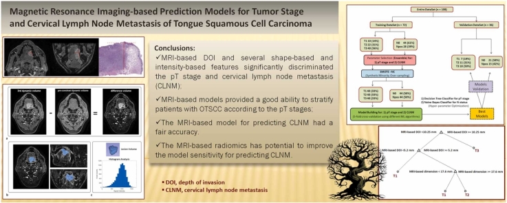
To evaluate the ability of preoperative MRI-based measurements to predict the pathological T (pT) stage and cervical lymph node metastasis (CLNM) via machine learning (ML)-driven models trained in oral tongue squamous cell carcinoma (OTSCC).
108 patients with a new diagnosis of OTSCC were enrolled. The preoperative MRI study included post-contrast high-resolution T1-weighted images acquired in all patients. MRI-based depth of invasion (DOI) and tumor dimension-together with shape-based and intensity-based features-were extracted from the lesion volume segmentation. The entire dataset was randomly divided into a training set and a validation set, and the performances of different types of ML algorithms were evaluated and compared.
MRI-based DOI and tumor dimension together with several shape-based and intensity-based signatures significantly discriminated the pT stage and LN status. The overall accuracy of the model for predicting the pT stage was 0.86 (95%CI, 0.78-0.92) and 0.81 (0.64-0.91) in the training and validation sets, respectively. There was no improvement in the model performance upon including shape-based and intensity-based features. The model for predicting CLNM based on DOI and tumor dimensions had a fair accuracy of 0.68 (0.57-0.78) and 0.69 (0.51-0.84) in the training and validation sets, respectively. The shape-based and intensity-based signatures have shown potential for improving the model sensitivity, with a comparable accuracy.
MRI-based models driven by ML algorithms could stratify patients with OTSCC according to the pT stages. They had a moderate ability to predict cervical lymph node metastasis.
通过在口腔舌鳞状细胞癌(OTSCC)中训练的机器学习(ML)驱动模型,评估基于术前MRI测量预测病理T(pT)分期和颈部淋巴结转移(CLNM)的能力。
纳入108例新诊断为OTSCC的患者。术前MRI检查包括所有患者的增强后高分辨率T1加权图像。从病变体积分割中提取基于MRI的浸润深度(DOI)和肿瘤尺寸,以及基于形状和基于强度的特征。将整个数据集随机分为训练集和验证集,并评估和比较不同类型ML算法的性能。
基于MRI的DOI和肿瘤尺寸以及一些基于形状和基于强度的特征显著区分了pT分期和淋巴结状态。预测pT分期模型在训练集和验证集中的总体准确率分别为0.86(95%CI,0.78-0.92)和0.81(0.64-0.91)。纳入基于形状和基于强度的特征后,模型性能没有改善。基于DOI和肿瘤尺寸预测CLNM的模型在训练集和验证集中的准确率分别为0.68(0.57-0.78)和0.69(0.51-0.84)。基于形状和基于强度的特征显示出提高模型敏感性的潜力,且准确率相当。
由ML算法驱动的基于MRI的模型可以根据pT分期对OTSCC患者进行分层。它们具有中等的预测颈部淋巴结转移的能力。