McNaughton Jake, Fernandez Justin, Holdsworth Samantha, Chong Benjamin, Shim Vickie, Wang Alan
Auckland Bioengineering Institute, University of Auckland, 6/70 Symonds Street, Auckland 1010, New Zealand.
Department of Engineering Science and Biomedical Engineering, University of Auckland, 3/70 Symonds Street, Auckland 1010, New Zealand.
Bioengineering (Basel). 2023 Sep 12;10(9):1078. doi: 10.3390/bioengineering10091078.
CT scans are often the first and only form of brain imaging that is performed to inform treatment plans for neurological patients due to its time- and cost-effective nature. However, MR images give a more detailed picture of tissue structure and characteristics and are more likely to pick up abnormalities and lesions. The purpose of this paper is to review studies which use deep learning methods to generate synthetic medical images of modalities such as MRI and CT.
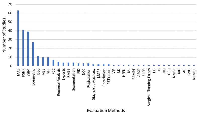
A literature search was performed in March 2023, and relevant articles were selected and analyzed. The year of publication, dataset size, input modality, synthesized modality, deep learning architecture, motivations, and evaluation methods were analyzed.
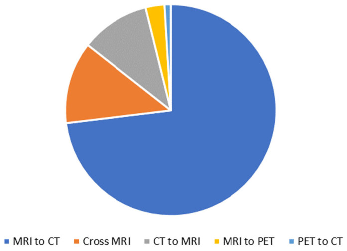
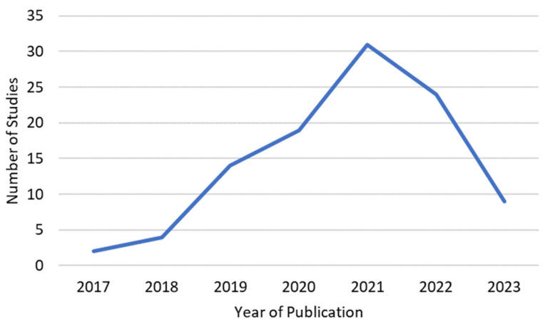
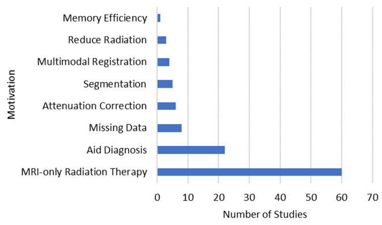
A total of 103 studies were included in this review, all of which were published since 2017. Of these, 74% of studies investigated MRI to CT synthesis, and the remaining studies investigated CT to MRI, Cross MRI, PET to CT, and MRI to PET. Additionally, 58% of studies were motivated by synthesizing CT scans from MRI to perform MRI-only radiation therapy. Other motivations included synthesizing scans to aid diagnosis and completing datasets by synthesizing missing scans.
Considerably more research has been carried out on MRI to CT synthesis, despite CT to MRI synthesis yielding specific benefits. A limitation on medical image synthesis is that medical datasets, especially paired datasets of different modalities, are lacking in size and availability; it is therefore recommended that a global consortium be developed to obtain and make available more datasets for use. Finally, it is recommended that work be carried out to establish all uses of the synthesis of medical scans in clinical practice and discover which evaluation methods are suitable for assessing the synthesized images for these needs.
由于CT扫描具有时间和成本效益,它通常是为神经科患者制定治疗方案时进行的首次也是唯一的脑成像检查方式。然而,磁共振成像(MR)图像能更详细地呈现组织结构和特征,更有可能发现异常和病变。本文的目的是综述使用深度学习方法生成诸如MRI和CT等模态的合成医学图像的研究。
于2023年3月进行文献检索,选择并分析相关文章。分析了发表年份、数据集大小、输入模态、合成模态、深度学习架构、动机和评估方法。
本综述共纳入103项研究,均自2017年以来发表。其中,74%的研究调查了从MRI到CT的合成,其余研究调查了从CT到MRI、交叉MRI、PET到CT以及MRI到PET。此外,58%的研究动机是从MRI合成CT扫描以进行仅基于MRI的放射治疗。其他动机包括合成扫描以辅助诊断以及通过合成缺失扫描来完善数据集。
尽管从CT到MRI的合成有其特定益处,但对从MRI到CT合成的研究要多得多。医学图像合成的一个限制是医学数据集,尤其是不同模态的配对数据集,在规模和可获取性方面都很欠缺;因此建议成立一个全球联盟来获取并提供更多可供使用的数据集。最后,建议开展工作以确立医学扫描合成在临床实践中的所有用途,并发现哪些评估方法适用于评估满足这些需求的合成图像。