• 文献检索
  • 文档翻译
  • 深度研究
  • 学术资讯
  • Suppr Zotero 插件Zotero 插件
  • 邀请有礼
  • 套餐&价格
  • 历史记录
应用&插件
Suppr Zotero 插件Zotero 插件浏览器插件Mac 客户端Windows 客户端微信小程序
定价
高级版会员购买积分包购买API积分包
服务
文献检索文档翻译深度研究API 文档MCP 服务
关于我们
关于 Suppr公司介绍联系我们用户协议隐私条款
关注我们

Suppr 超能文献

核心技术专利:CN118964589B侵权必究
粤ICP备2023148730 号-1Suppr @ 2026

文献检索

告别复杂PubMed语法,用中文像聊天一样搜索,搜遍4000万医学文献。AI智能推荐,让科研检索更轻松。

立即免费搜索

文件翻译

保留排版,准确专业,支持PDF/Word/PPT等文件格式,支持 12+语言互译。

免费翻译文档

深度研究

AI帮你快速写综述,25分钟生成高质量综述,智能提取关键信息,辅助科研写作。

立即免费体验

硅酸钙骨水泥中pH值及钙离子释放的评估:一项体外对比研究

Assessment of pH Value and Release of Calcium Ions in Calcium Silicate Cements: An In Vitro Comparative Study.

作者信息

Herrera-Trinidad Rubén, Molinero-Mourelle Pedro, Fonseca Manrique, Weber Adrian Roman, Vera Vicente, Mena María Luz, Vera-González Vicente

机构信息

Department of Conservative Dentistry and Orofacial Prosthodontics, Faculty of Dentistry, Complutense University of Madrid, 28040 Madrid, Spain.

Department of Reconstructive Dentistry and Gerodontology, School of Dental Medicine, University of Bern, 3007 Bern, Switzerland.

出版信息

Materials (Basel). 2023 Sep 14;16(18):6213. doi: 10.3390/ma16186213.

DOI:10.3390/ma16186213
PMID:37763492
原文链接:https://pmc.ncbi.nlm.nih.gov/articles/PMC10532804/
Abstract

UNLABELLED

The goal of this study was to evaluate the pH and the release of calcium from four calcium-silicate-based cements.

METHODS

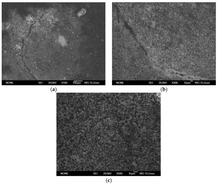
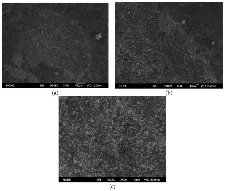
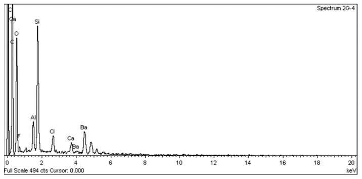
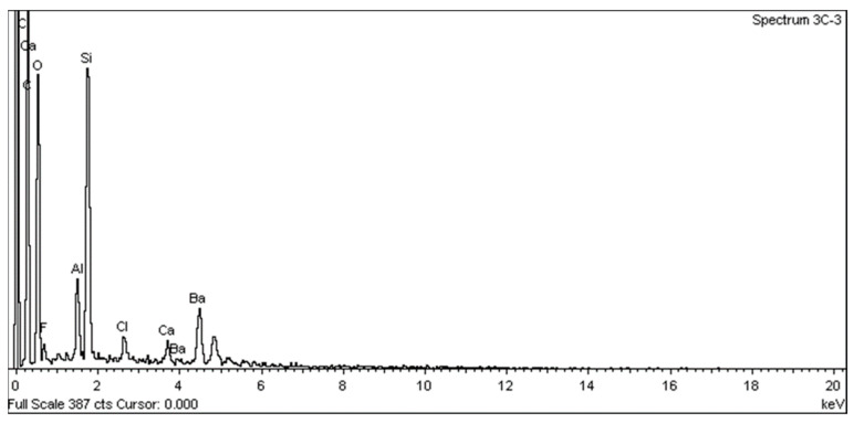
Four materials were tested (ProClinic MTA; Angelus MTA; ProRoot MTA; Biodentine). The palatal canal root of acrylic upper molars was filled with each cement. Afterwards, they were set in phosphate-buffered saline. Measurements were taken by atomic adsorption spectroscopy (AAS) at 3, 24, 72, 168, 336, 672, and 1008 h. The pH was measured at the same timepoints. Kruskal-Wallis tests were carried out in each period, as the Kolmogorov-Smirnov and Shapiro-Wilk tests showed no parametric results.

RESULTS

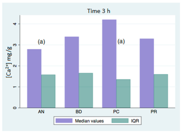
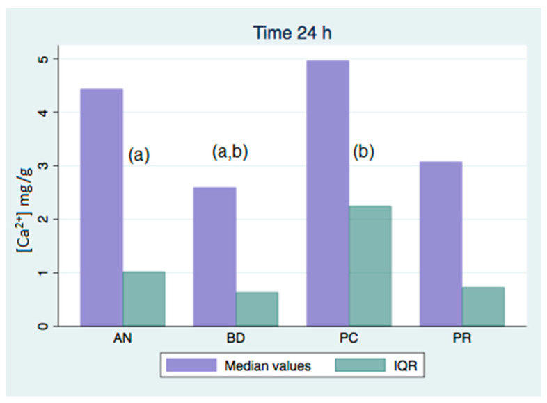
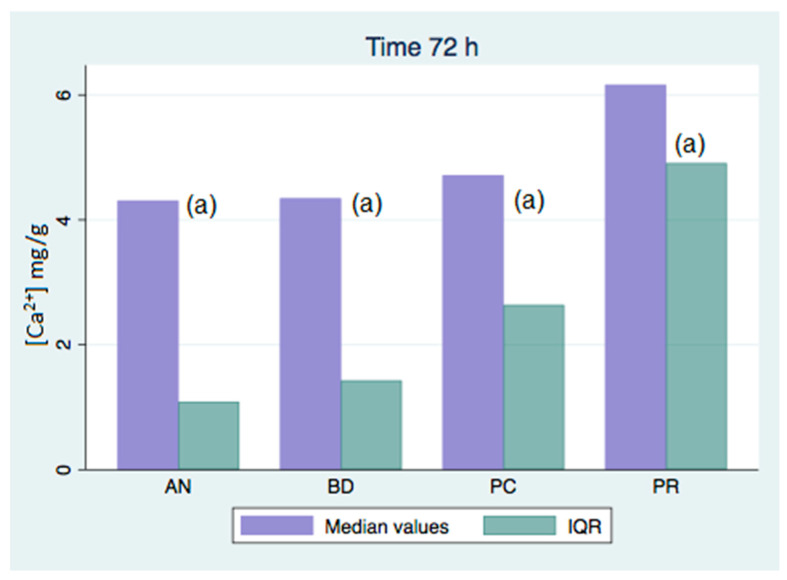
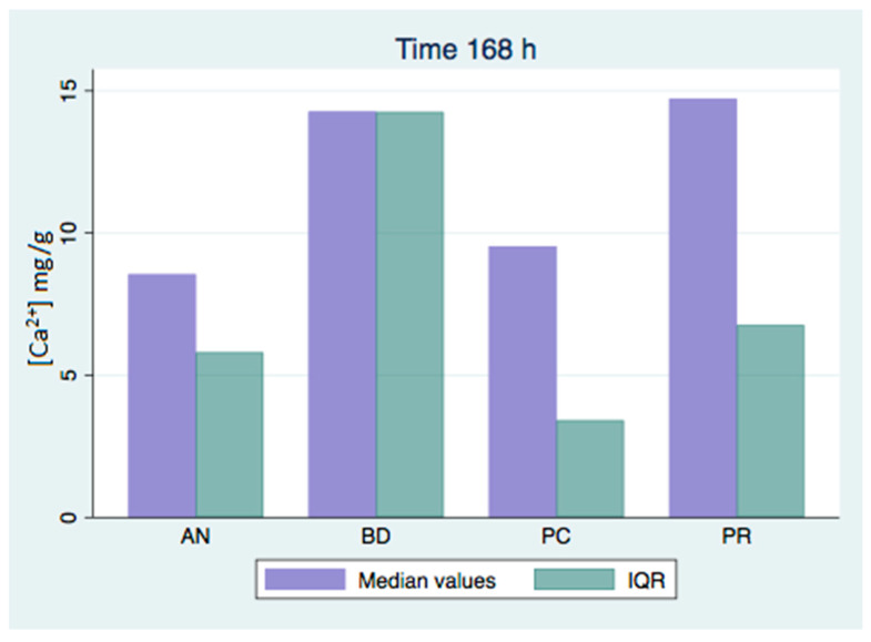
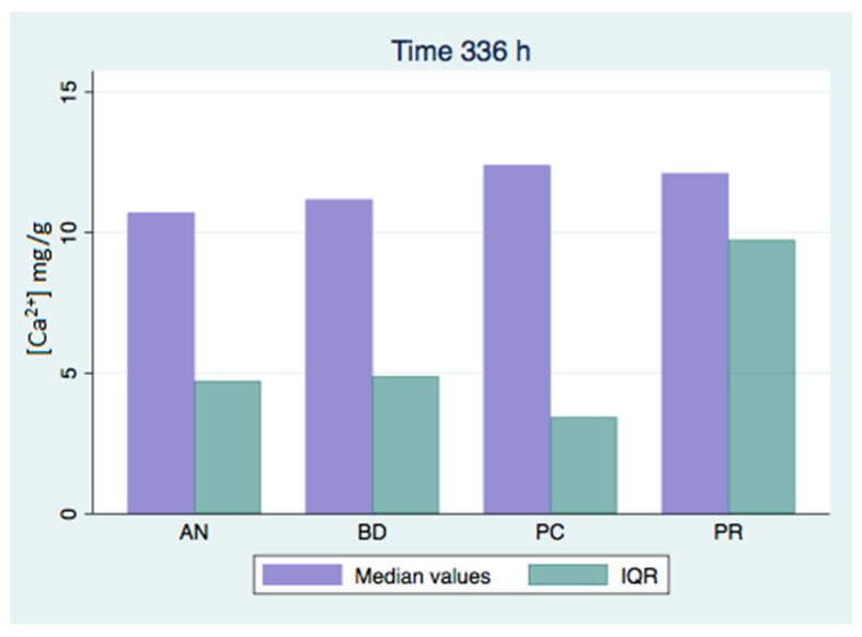
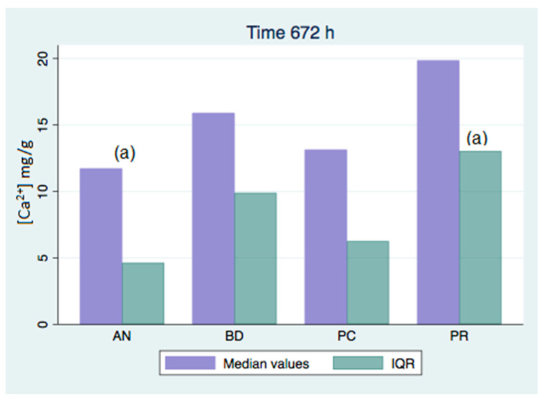
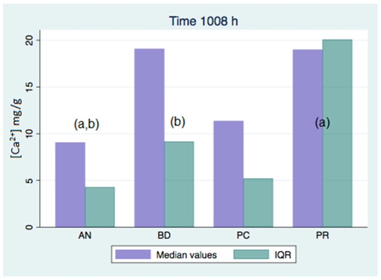
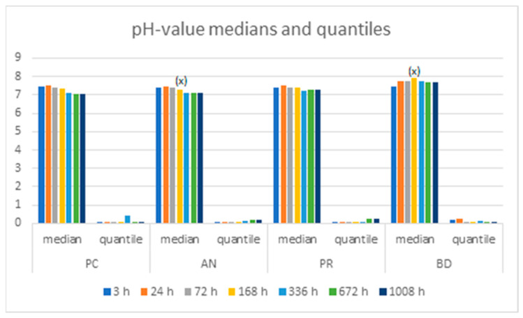
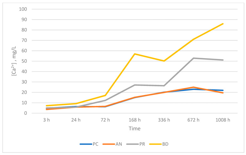
Significant differences ( < 0.05) in calcium release were found at the 3-, 24-, and 72-hour evaluations. All of the analyzed groups presented a release of calcium ions up to 168 h, and the general tendency was to increase up to 672 h, with a maximum release of 25.45 mg/g in the ProRoot group. We could only observe significant differences ( < 0.05) in pH value over 168 h between the Biodentine (7.93) and Angelus MTA (7.31) groups.

CONCLUSIONS

There were significant differences ( < 0.05) in calcium release. Nevertheless, no significant differences ( > 0.05) in the pH values were found at the studied timepoints, except for the values at 168 h.

摘要

未标注

本研究的目的是评估四种硅酸钙基水门汀的pH值和钙释放情况。

方法

测试了四种材料(ProClinic MTA;Angelus MTA;ProRoot MTA;Biodentine)。用每种水门汀填充丙烯酸上磨牙的腭根管。之后,将它们置于磷酸盐缓冲盐溶液中。在3、24、72、168、336、672和1008小时通过原子吸收光谱法(AAS)进行测量。在相同时间点测量pH值。由于Kolmogorov-Smirnov和Shapiro-Wilk检验未显示参数结果,因此在每个时间段进行Kruskal-Wallis检验。

结果

在3小时、24小时和72小时的评估中发现钙释放存在显著差异(<0.05)。所有分析组在168小时内均呈现钙离子释放,总体趋势是在672小时内增加,ProRoot组的最大释放量为25.45mg/g。我们仅观察到Biodentine组(7.93)和Angelus MTA组(7.31)在168小时以上的pH值存在显著差异(<0.05)。

结论

钙释放存在显著差异(<0.05)。然而,除了168小时的值外,在所研究的时间点pH值未发现显著差异(>0.05)。

https://cdn.ncbi.nlm.nih.gov/pmc/blobs/4ade/10532804/f264c1fb286c/materials-16-06213-g018.jpg
https://cdn.ncbi.nlm.nih.gov/pmc/blobs/4ade/10532804/e0c30a3e17ec/materials-16-06213-g001.jpg
https://cdn.ncbi.nlm.nih.gov/pmc/blobs/4ade/10532804/4e6046ce4414/materials-16-06213-g002.jpg
https://cdn.ncbi.nlm.nih.gov/pmc/blobs/4ade/10532804/6d07c27132b9/materials-16-06213-g003.jpg
https://cdn.ncbi.nlm.nih.gov/pmc/blobs/4ade/10532804/24e34d87300d/materials-16-06213-g004.jpg
https://cdn.ncbi.nlm.nih.gov/pmc/blobs/4ade/10532804/cf0c6195987d/materials-16-06213-g005.jpg
https://cdn.ncbi.nlm.nih.gov/pmc/blobs/4ade/10532804/a6c4c55eae28/materials-16-06213-g006.jpg
https://cdn.ncbi.nlm.nih.gov/pmc/blobs/4ade/10532804/46efa23546a2/materials-16-06213-g007.jpg
https://cdn.ncbi.nlm.nih.gov/pmc/blobs/4ade/10532804/eb49106c5761/materials-16-06213-g008.jpg
https://cdn.ncbi.nlm.nih.gov/pmc/blobs/4ade/10532804/db0619e1ef57/materials-16-06213-g009.jpg
https://cdn.ncbi.nlm.nih.gov/pmc/blobs/4ade/10532804/6e6cb8361551/materials-16-06213-g010.jpg
https://cdn.ncbi.nlm.nih.gov/pmc/blobs/4ade/10532804/630e2ed061f2/materials-16-06213-g011.jpg
https://cdn.ncbi.nlm.nih.gov/pmc/blobs/4ade/10532804/25c7d8c6026e/materials-16-06213-g012.jpg
https://cdn.ncbi.nlm.nih.gov/pmc/blobs/4ade/10532804/77636e2e4549/materials-16-06213-g013.jpg
https://cdn.ncbi.nlm.nih.gov/pmc/blobs/4ade/10532804/b209fe0b030f/materials-16-06213-g014.jpg
https://cdn.ncbi.nlm.nih.gov/pmc/blobs/4ade/10532804/8f2e713d181d/materials-16-06213-g015.jpg
https://cdn.ncbi.nlm.nih.gov/pmc/blobs/4ade/10532804/24e6aa19f0e4/materials-16-06213-g016.jpg
https://cdn.ncbi.nlm.nih.gov/pmc/blobs/4ade/10532804/e6a9fd308f5e/materials-16-06213-g017.jpg
https://cdn.ncbi.nlm.nih.gov/pmc/blobs/4ade/10532804/f264c1fb286c/materials-16-06213-g018.jpg

相似文献

1
Assessment of pH Value and Release of Calcium Ions in Calcium Silicate Cements: An In Vitro Comparative Study.硅酸钙骨水泥中pH值及钙离子释放的评估:一项体外对比研究
Materials (Basel). 2023 Sep 14;16(18):6213. doi: 10.3390/ma16186213.
2
Biointeractivity-related versus chemi/physisorption-related apatite precursor-forming ability of current root end filling materials.当前根管充填材料的生物相互作用相关与化学/物理吸附相关的磷灰石前体形成能力。
J Biomed Mater Res B Appl Biomater. 2013 Oct;101(7):1107-23. doi: 10.1002/jbm.b.32920. Epub 2013 Apr 4.
3
Influence of composition on setting kinetics of new injectable and/or fast setting tricalcium silicate cements.成分对新型可注射和/或快凝硅酸三钙水泥凝结动力学的影响。
Dent Mater. 2014 Dec;30(12):1291-303. doi: 10.1016/j.dental.2014.09.005. Epub 2014 Oct 23.
4
Ion release and mechanical properties of calcium silicate and calcium hydroxide materials used for pulp capping.用于盖髓的硅酸钙和氢氧化钙材料的离子释放和机械性能。
Int Endod J. 2015 Jan;48(1):89-94. doi: 10.1111/iej.12281. Epub 2014 Apr 18.
5
The physical properties and ion release of CPP-ACP-modified calcium silicate-based cements.CPP-ACP 改性硅酸钙基水泥的物理性能及离子释放
Aust Dent J. 2015 Dec;60(4):434-44. doi: 10.1111/adj.12255.
6
Effect of acid etching procedures on the compressive strength of 4 calcium silicate-based endodontic cements.酸蚀程序对4种硅酸钙基根管封闭剂抗压强度的影响
J Endod. 2013 Dec;39(12):1646-8. doi: 10.1016/j.joen.2013.09.008. Epub 2013 Oct 15.
7
Evaluation of the physical and chemical properties of two commercial and three experimental root-end filling materials.两种市售和三种实验性根尖充填材料的物理和化学性质评估。
Oral Surg Oral Med Oral Pathol Oral Radiol Endod. 2010 Aug;110(2):250-6. doi: 10.1016/j.tripleo.2010.04.021.
8
Microstructure and chemical analysis of four calcium silicate-based cements in different environmental conditions.四种不同环境条件下硅酸钙基水泥的微观结构和化学分析。
Clin Oral Investig. 2019 Jan;23(1):43-52. doi: 10.1007/s00784-018-2394-1. Epub 2018 Mar 30.
9
Investigation of the hydration and bioactivity of radiopacified tricalcium silicate cement, Biodentine and MTA Angelus.研究放射显影硅酸三钙水泥、Biodentine 和 MTA Angelus 的水合和生物活性。
Dent Mater. 2013 May;29(5):580-93. doi: 10.1016/j.dental.2013.03.007. Epub 2013 Mar 26.
10
Six-Month Color Stability Assessment of Two Calcium Silicate-Based Cements Used in Regenerative Endodontic Procedures.用于再生牙髓治疗的两种硅酸钙基水门汀的六个月颜色稳定性评估
J Funct Biomater. 2019 Feb 28;10(1):14. doi: 10.3390/jfb10010014.

引用本文的文献

1
Clinical and radiographic evaluation of premixed bioceramic putty as an apical plug in nonvital immature anterior permanent teeth.预混生物陶瓷糊剂作为非活髓未成熟恒前牙根尖充填材料的临床及影像学评估
Sci Rep. 2025 Jul 21;15(1):26487. doi: 10.1038/s41598-025-11407-x.
2
Evaluation of the Effects of Retro-Cavity Preconditioning with or Without Ethylenediaminetetraacetic Acid on Root Surface pH and Dislodgement Resistance of NeoMTA2 and Mineral Trioxide Aggregate Flow Retro-Fills: An Ex Vivo Investigation.评估含或不含乙二胺四乙酸的根管内预处理对NeoMTA2和流动型三氧化矿物凝聚体根管倒充填物的根面pH值及抗脱位性的影响:一项体外研究
J Funct Biomater. 2024 Dec 24;16(1):3. doi: 10.3390/jfb16010003.
3

本文引用的文献

1
Biocompatibility and mineralization potential of new calcium silicate cements.新型硅酸钙水门汀的生物相容性及矿化潜力
J Dent Sci. 2023 Jul;18(3):1189-1198. doi: 10.1016/j.jds.2022.10.004. Epub 2022 Oct 20.
2
New Bioactive Calcium Silicate Cement Mineral Trioxide Aggregate Repair High Plasticity (MTA HP)-A Systematic Review.新型生物活性硅酸钙水泥三氧化物矿物聚合体修复高可塑性材料(MTA HP)——系统评价
Materials (Basel). 2021 Aug 14;14(16):4573. doi: 10.3390/ma14164573.
3
A Microstructure Insight of MTA Repair HP of Rapid Setting Capacity and Bioactive Response.
Enhancing pH Modulation and Calcium Ions Release in External Resorption Artificial Defects: A Comparative Analysis of Ultrasonic Activation Effects on Ca(OH), MTA, and CEM Cement.
增强外部吸收人工缺损中的pH调节和钙离子释放:超声激活对Ca(OH)₂、MTA和CEM水泥影响的比较分析
Int J Dent. 2024 Nov 14;2024:8850548. doi: 10.1155/2024/8850548. eCollection 2024.
4
Different pulp capping agents and their effect on pulp inflammatory response: A narrative review.不同的盖髓剂及其对牙髓炎症反应的影响:一项叙述性综述。
Saudi Dent J. 2024 Oct;36(10):1295-1306. doi: 10.1016/j.sdentj.2024.07.014. Epub 2024 Jul 30.
5
Evaluation of biocompatibility and bioactive potential of Well-Root PT by comparison with ProRoot MTA and Biodentine.通过与ProRoot MTA和Biodentine比较评估Well-Root PT的生物相容性和生物活性潜力。
J Dent Sci. 2024 Oct;19(4):2218-2225. doi: 10.1016/j.jds.2024.03.004. Epub 2024 Mar 11.
6
Comparative Analysis of Calcium and Hydroxyl Ion Release from Pulp Dressing Materials in Pulpotomized Premolars.比较分析在活髓切断术前磨牙中牙髓敷料材料的钙离子和氢氧根离子释放。
Med Sci Monit. 2024 Sep 28;30:e945089. doi: 10.12659/MSM.945089.
具有快速凝固能力和生物活性反应的MTA修复HP的微观结构洞察
Materials (Basel). 2020 Apr 2;13(7):1641. doi: 10.3390/ma13071641.
4
Release of Transforming Growth Factor Beta 1 from Human Tooth Dentin after Application of Either ProRoot MTA or Biodentine as a Coronal Barrier.ProRoot MTA 或 Biodentine 用作冠方屏障后,人牙本质中转化生长因子 β1 的释放。
J Endod. 2019 Jun;45(6):701-705. doi: 10.1016/j.joen.2019.03.011. Epub 2019 Apr 25.
5
How far do calcium release measurements properly reflect its multiple roles in dental tissue mineralization?钙释放测量在多大程度上能准确反映其在牙齿组织矿化中的多种作用?
Clin Oral Investig. 2019 Jan;23(1):501. doi: 10.1007/s00784-018-2789-z. Epub 2019 Jan 5.
6
Calcium release and physical properties of modified carbonate apatite cement as pulp capping agent in dental application.改性碳酸磷灰石水泥作为牙科应用中盖髓剂的钙释放及物理性能
Biomater Res. 2018 Dec 6;22:35. doi: 10.1186/s40824-018-0146-6. eCollection 2018.
7
Pulp capping materials modulate the balance between inflammation and regeneration.牙髓覆盖材料可调节炎症和再生之间的平衡。
Dent Mater. 2019 Jan;35(1):24-35. doi: 10.1016/j.dental.2018.09.008. Epub 2018 Sep 27.
8
Tricalcium Silicate Capping Materials Modulate Pulp Healing and Inflammatory Activity In Vitro.硅酸三钙盖髓材料对体外牙髓愈合和炎症活性的调节作用。
J Endod. 2018 Nov;44(11):1686-1691. doi: 10.1016/j.joen.2018.06.009. Epub 2018 Sep 11.
9
In vivo biocompatibility and biomineralization of calcium silicate cements.硅酸钙骨水泥的体内生物相容性与生物矿化
Eur J Oral Sci. 2018 Aug;126(4):326-333. doi: 10.1111/eos.12539. Epub 2018 Jul 1.
10
Effect of Exposed Surface Area, Volume and Environmental pH on the Calcium Ion Release of Three Commercially Available Tricalcium Silicate Based Dental Cements.暴露表面积、体积和环境pH值对三种市售硅酸三钙基牙科水门汀钙离子释放的影响
Materials (Basel). 2018 Jan 13;11(1):123. doi: 10.3390/ma11010123.