Yang Ying, Chen Huan, Zhang Caisheng, Shin Hyun-Jin, Qian Yingjuan, Jung Yong-Sam
One Health Laboratory, Jiangsu Foreign Expert Workstation, College of Veterinary Medicine, Nanjing Agricultural University, Nanjing 210095, China.
MOE Joint International Research Laboratory of Animal Health and Food Safety, College of Veterinary Medicine, Nanjing Agricultural University, Nanjing 210095, China.
Viruses. 2023 Sep 4;15(9):1874. doi: 10.3390/v15091874.
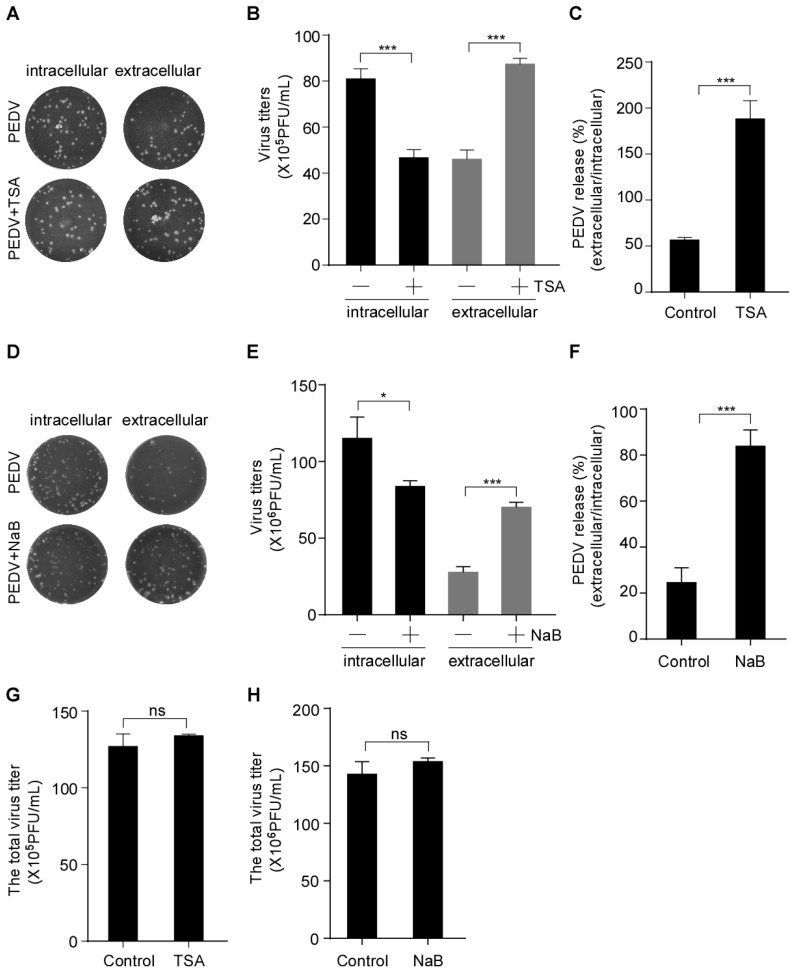
Porcine epidemic diarrhea virus (PEDV) is an alpha-coronavirus causing acute diarrhea and high mortality in neonatal suckling piglets, resulting in huge economic losses for the global swine industry. The replication, assembly and cell egression of PEDV, an enveloped RNA virus, are mediated via altered intracellular trafficking. The underlying mechanisms of PEDV secretion are poorly understood. In this study, we found that the histone deacetylase (HDAC)-specific inhibitors, trichostatin A (TSA) and sodium butyrate (NaB), facilitate the secretion of infectious PEDV particles without interfering with its assembly. We found that PEDV N protein and its replicative intermediate dsRNA colocalize with coat protein complex II (COPII)-coated vesicles. We also showed that the colocalization of PEDV and COPII is enhanced by the HDAC-specific inhibitors. In addition, ultrastructural analysis revealed that the HDAC-specific inhibitors promote COPII-coated vesicles carrying PEDV virions and the secretion of COPII-coated vesicles. Consistently, HDAC-specific inhibitors-induced PEDV particle secretion was abolished by Sec24B knockdown, implying that the HDAC-specific inhibitors-mediated COPII-coated vesicles are required for PEDV secretion. Taken together, our findings provide initial evidence suggesting that PEDV virions can assemble in the endoplasmic reticulum (ER) and bud off from the ER in the COPII-coated vesicles. HDAC-specific inhibitors promote PEDV release by hijacking the COPII-coated vesicles.
猪流行性腹泻病毒(PEDV)是一种α冠状病毒,可导致新生仔猪急性腹泻和高死亡率,给全球养猪业造成巨大经济损失。PEDV是一种包膜RNA病毒,其复制、组装和细胞出芽过程是通过改变细胞内运输来介导的。PEDV分泌的潜在机制尚不清楚。在本研究中,我们发现组蛋白去乙酰化酶(HDAC)特异性抑制剂曲古抑菌素A(TSA)和丁酸钠(NaB)可促进感染性PEDV颗粒的分泌,而不干扰其组装。我们发现PEDV N蛋白及其复制中间体双链RNA与II型被膜小泡(COPII)共定位。我们还表明,HDAC特异性抑制剂可增强PEDV与COPII的共定位。此外,超微结构分析显示,HDAC特异性抑制剂可促进携带PEDV病毒粒子的COPII被膜小泡以及COPII被膜小泡的分泌。一致的是,Sec24B基因敲低消除了HDAC特异性抑制剂诱导的PEDV颗粒分泌,这意味着HDAC特异性抑制剂介导的COPII被膜小泡是PEDV分泌所必需的。综上所述,我们的研究结果提供了初步证据,表明PEDV病毒粒子可在内质网(ER)中组装,并从ER中以COPII被膜小泡的形式出芽。HDAC特异性抑制剂通过劫持COPII被膜小泡促进PEDV释放。