Translational Medical Center for Stem Cell Therapy and Institutes for Regenerative Medicine, Shanghai East Hospital, Tongji University School of Medicine, 1800 Yuntai Rd., Shanghai, 200123, China.
Department of Cardiovascular and Thoracic Surgery, Shanghai East Hospital, Tongji University School of Medicine, Shanghai, 200120, China.
Stem Cell Res Ther. 2023 Sep 29;14(1):278. doi: 10.1186/s13287-023-03462-w.
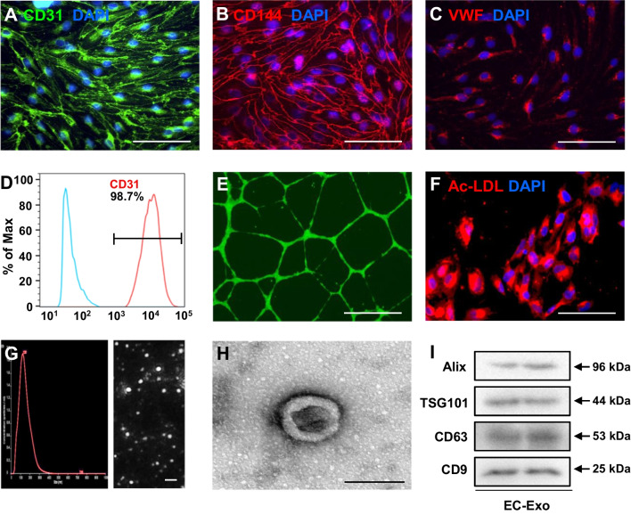
Human induced pluripotent stem cell-derived endothelial cells (hiPSC-ECs) exhibit the potential to repair the injured heart after myocardial infarction (MI) by promoting neovascularization and cardiomyocyte survival. However, because of the low cellular retention and poor engraftment efficacy, cell therapy of MI is partly mediated by exosomes secreted from the transplanted cells. In this study, we investigated whether exosomes secreted from hiPSC-ECs could become a promising acellular approach to repair the infarcted heart after MI and elucidated the underlying protective mechanism.
The hiPSC-ECs were differentiated, and exosomes were isolated in vitro. Then, hiPSC-EC exosomes were delivered by intramyocardial injection in a murine MI model in vivo. Echocardiography, combined with hemodynamic measurement, histological examination, Ca transient and cell shortening assessment, and Western blot, was used to determine the protective effects of hiPSC-EC exosomes on the infarcted heart. Furthermore, microRNA sequencing, luciferase activity assay, and microRNA gain-loss function experiments were performed to investigate the enriched microRNA and its role in exosome-mediated effects.
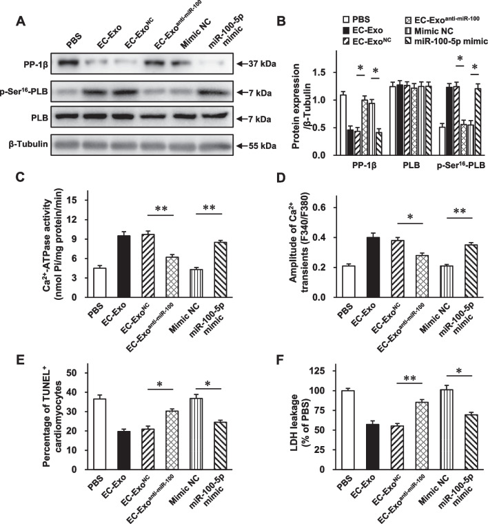
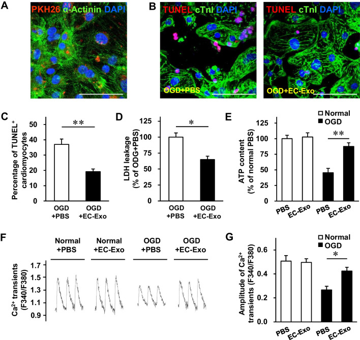
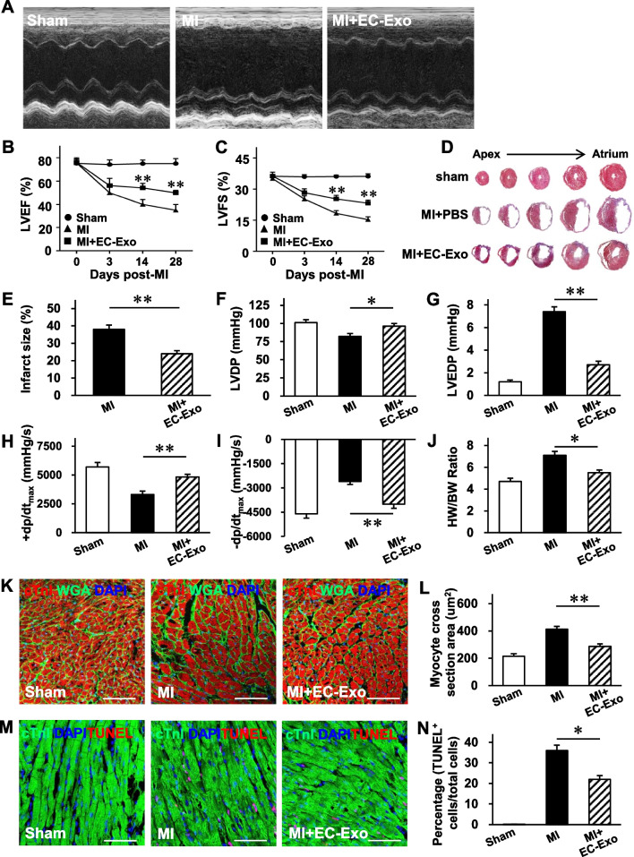
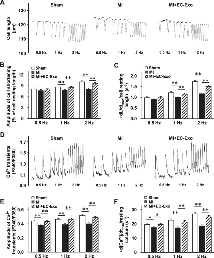
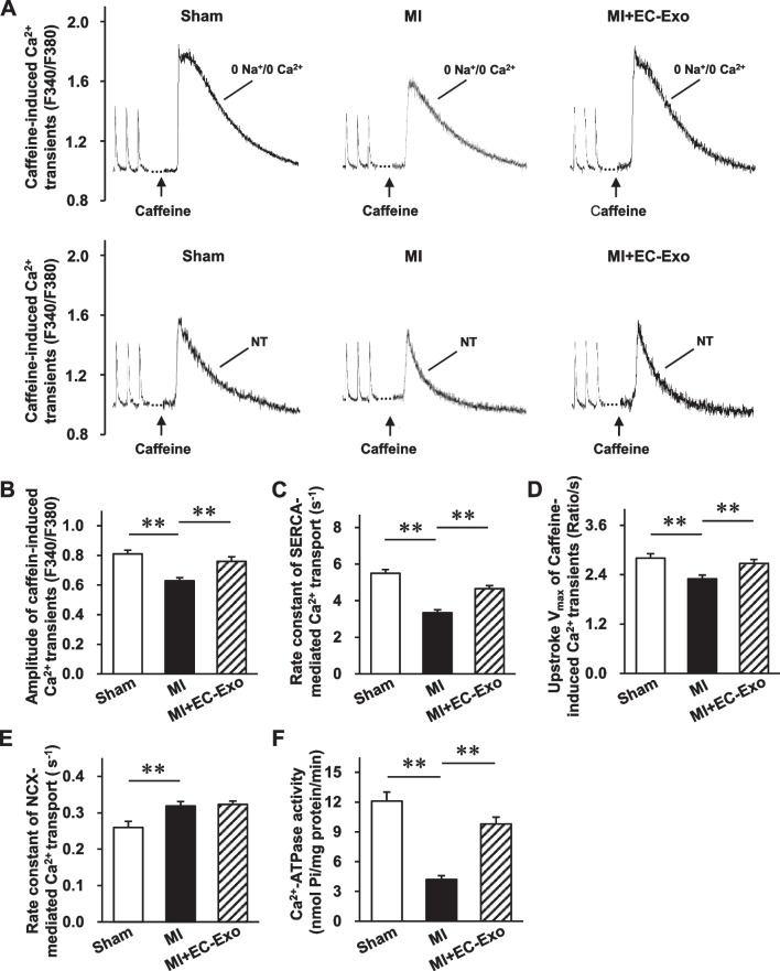
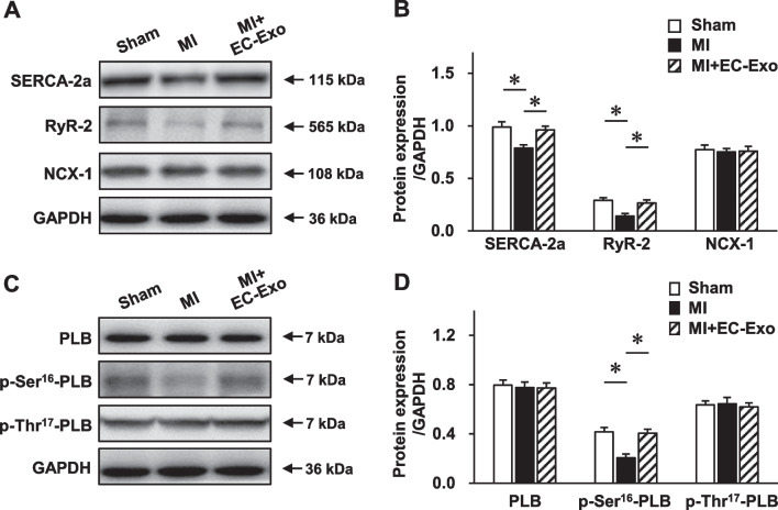
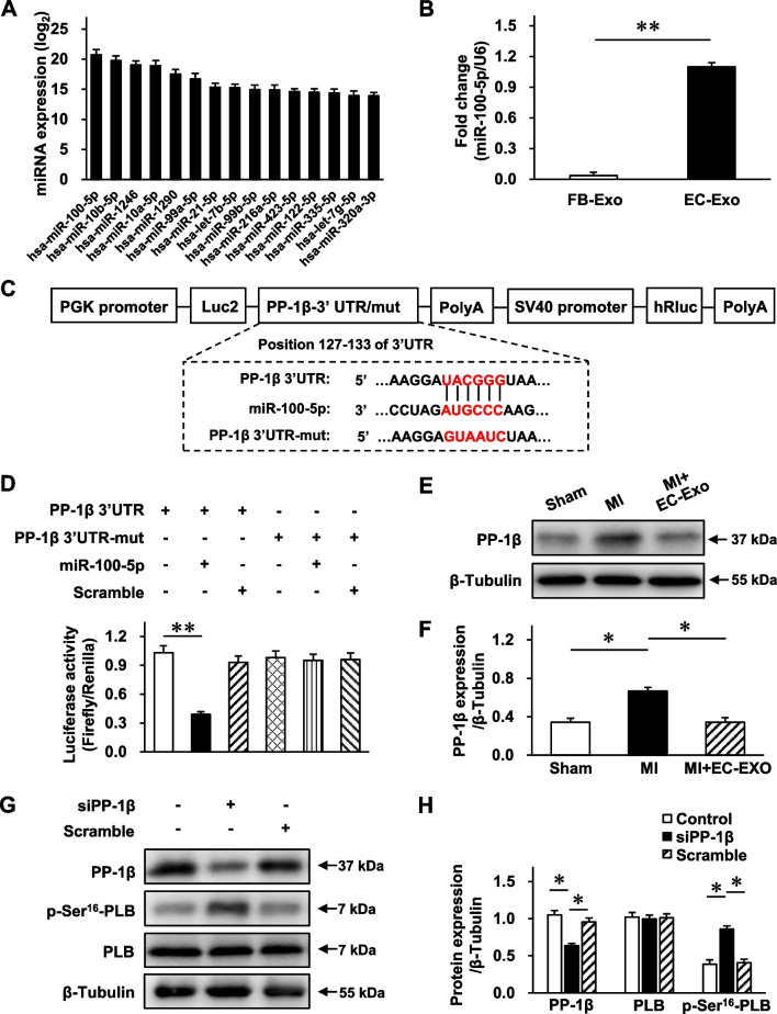
In vitro, the hiPSC-EC exosomes enhanced intracellular Ca transients, increased ATP content, and improved cell survival to protect cardiomyocytes from oxygen-glucose deprivation injury. Congruously, hiPSC-EC exosome administration in vivo improved the myocardial contractile function and attenuated the harmful left ventricular remodeling after MI without increasing the frequency of arrhythmias. Mechanistically, the hiPSC-EC exosomes notably rescued the protein expression and function of the sarcoplasmic reticulum Ca ATPase 2a (SERCA-2a) and ryanodine receptor 2 (RyR-2) to maintain intracellular Ca homeostasis and increase cardiomyocyte contraction after MI. The microRNA sequencing showed that miR-100-5p was the most abundant microRNA in exosomes. miR-100-5p could target protein phosphatase 1β (PP-1β) to enhance phospholamban (PLB) phosphorylation at Ser and subsequent SERCA activity, which contributes to the hiPSC-EC exosome-exerted cytoprotective effects on maintaining intracellular Ca homeostasis and promoting cardiomyocyte survival.
The hiPSC-EC exosomes maintain cardiomyocyte Ca homeostasis to improve myocardial recovery after MI, which may provide an acellular therapeutic option for myocardial injury.
人诱导多能干细胞衍生的内皮细胞(hiPSC-ECs)通过促进血管生成和心肌细胞存活,具有修复心肌梗死后受损心脏的潜力。然而,由于细胞保留率低和移植细胞的植入效果差,细胞疗法部分通过移植细胞分泌的外泌体介导。在这项研究中,我们研究了 hiPSC-EC 分泌的外泌体是否可以成为一种有前途的无细胞方法来修复心肌梗死后的受损心脏,并阐明了潜在的保护机制。
hiPSC-EC 在体外分化,然后在体外分离外泌体。然后,将 hiPSC-EC 外泌体通过心肌内注射递送至体内的小鼠心肌梗死模型中。通过超声心动图、血流动力学测量、组织学检查、钙瞬变和细胞缩短评估以及 Western blot,确定 hiPSC-EC 外泌体对梗死心脏的保护作用。此外,进行 microRNA 测序、荧光素酶活性测定和 microRNA 增益-损耗功能实验,以研究富含 microRNA 及其在 exosome 介导作用中的作用。
在体外,hiPSC-EC 外泌体增强了细胞内钙瞬变,增加了 ATP 含量,并改善了细胞存活,从而保护心肌细胞免受氧葡萄糖剥夺损伤。一致地,hiPSC-EC 外泌体的体内给药改善了心肌收缩功能,并减轻了心肌梗死后的有害左室重构,而不会增加心律失常的频率。在机制上,hiPSC-EC 外泌体显著挽救了肌浆网 Ca2+-ATP 酶 2a(SERCA-2a)和 Ryanodine 受体 2(RyR-2)的蛋白表达和功能,以维持细胞内 Ca2+稳态,并增加心肌梗死后的心肌收缩。microRNA 测序表明,miR-100-5p 是外泌体中最丰富的 microRNA。miR-100-5p 可以靶向蛋白磷酸酶 1β(PP-1β),以增强肌球蛋白轻链磷酸酶(PLB)丝氨酸磷酸化和随后的 SERCA 活性,这有助于 hiPSC-EC 外泌体对维持细胞内 Ca2+稳态和促进心肌细胞存活的细胞保护作用。
hiPSC-EC 外泌体维持心肌细胞 Ca2+稳态,改善心肌梗死后的心肌恢复,可为心肌损伤提供一种无细胞治疗选择。