Department of Orthopaedic Surgery, CHA Bundang Medical Center, CHA University School of Medicine, Gyeonggi-do, Republic of Korea.
SL Bio, Inc., Gyeonggi-do, Republic of Korea.
Front Endocrinol (Lausanne). 2023 Sep 19;14:1238654. doi: 10.3389/fendo.2023.1238654. eCollection 2023.
Osteoporotic vertebral compression fractures commonly involve the superior vertebral body; however, their associated causes have not yet been clearly established. This study aimed to determine the trabecular structural differences between the superior and inferior regions of the vertebral body using cadaveric and clinical studies.
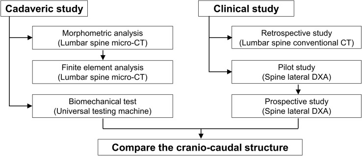
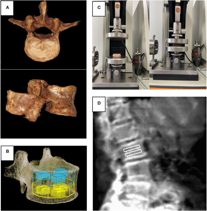
First, five vertebrae were collected from three human cadavers. The trabecular structures of the superior and inferior regions of each vertebral body were analyzed using micro-computed tomography (micro-CT), finite element analysis (FEA), and biomechanical test. Based on the results of the ex vivo study, we conducted a clinical study. Second, spine CT images were retrospectively collected. Bone volume and Hounsfield unit were analyzed for 192 vertebral bodies. Finally, after sample size calculation based on the pilot study, prospectively, 200 participants underwent dual-energy X-ray absorptiometry (DXA) of the lateral spine. The bone mineral densities (BMDs) of the superior and inferior regions of each lumbar vertebral body were measured. The paired t-test and Wilcoxon signed-rank test were used for the statistical analyses, and p-value < 0.05 was considered significant.
Cadaver studies revealed differences between the superior and inferior trabecular bone structures. The bone volume ratio, BMD, and various other trabecular parameters advocated for decreased strength of the superior region. Throughout the biomechanical study, the limitations of the compression force were 3.44 and 4.63 N/m for the superior and inferior regions, respectively. In the FEA study, the inferior region had a lower average displacement and higher von Mises stress than the superior region. In the clinical spine CT-based bone volume and BMD study, the bone volume was significantly higher in the inferior region than in the superior region. In the lateral spine DXA, the mean BMD of the superior region of vertebral bodies was significantly lower compared with that of the inferior region.
The superior trabecular structure of the lumbar vertebral bodies possesses more biomechanical susceptibility compared with the inferior trabecular structure, confirming its dominant role in causing osteoporotic vertebral fractures. Physicians should also focus on the BMD values of the superior region of the vertebral body using lateral spine DXA to evaluate osteoporosis.
骨质疏松性椎体压缩骨折常累及椎体上部;然而,其相关原因尚未明确。本研究旨在通过尸体和临床研究来确定椎体上下部的小梁结构差异。
首先,从三具人体尸体中收集了五节椎体。使用微计算机断层扫描(micro-CT)、有限元分析(FEA)和生物力学测试分析每个椎体上下部的小梁结构。基于体外研究结果,我们进行了一项临床研究。其次,回顾性地收集脊柱 CT 图像。分析了 192 个椎体的骨体积和 Hounsfield 单位。最后,根据初步研究进行样本量计算后,前瞻性地对 200 名参与者进行侧位脊柱双能 X 线吸收法(DXA)检查。测量每个腰椎椎体上下部的骨密度(BMD)。采用配对 t 检验和 Wilcoxon 符号秩检验进行统计学分析,p 值<0.05 认为有统计学意义。
尸体研究显示椎体上下部小梁骨结构存在差异。骨体积比、BMD 和其他各种小梁参数表明,上区域的强度降低。在整个生物力学研究中,上下区域的压缩力限制分别为 3.44 和 4.63 N/m。在 FEA 研究中,下区域的平均位移较低,von Mises 应力较高。在基于临床脊柱 CT 的骨体积和 BMD 研究中,下区域的骨体积明显高于上区域。在侧位脊柱 DXA 中,与下区域相比,椎体上区域的平均 BMD 明显较低。
与下区域的小梁结构相比,腰椎体上区域的小梁结构具有更高的生物力学易感性,这证实了其在导致骨质疏松性椎体骨折方面的主导作用。医生还应使用侧位脊柱 DXA 关注椎体上区域的 BMD 值,以评估骨质疏松症。