Division of Epigenomics, National Cancer Center Research Institute, Tokyo, Japan.
Division of Surgery, University of Kyoto, Kyoto, Japan.
PLoS One. 2023 Oct 5;18(10):e0290034. doi: 10.1371/journal.pone.0290034. eCollection 2023.
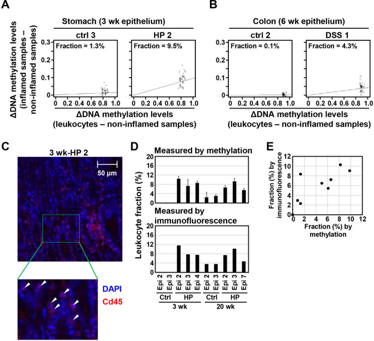
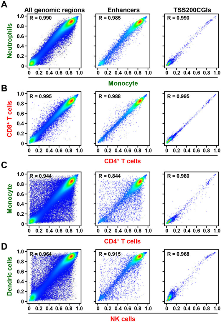
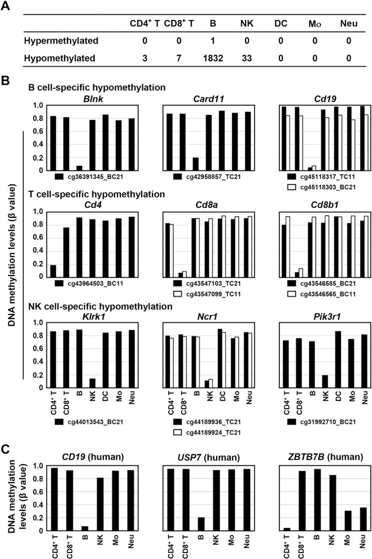
Precise analysis of tissue DNA and RNA samples is often hampered by contaminating non-target cells whose amounts are highly variable. DNA methylation profiles are specific to cell types, and can be utilized for assessment of the fraction of such contaminating non-target cells. Here, we aimed 1) to identify methylation profiles specific to multiple types of mouse leukocytes, and 2) to estimate the fraction of leukocytes infiltrating inflamed tissues using DNA samples. First, genome-wide DNA methylation analysis was conducted for three myeloid-lineage cells and four lymphoid-lineage cells isolated by fluorescence-activated cell sorting after magnetic-activated cell sorting from leukocytes in the spleen. Clustering analysis using CpG sites within enhancers separated the three myeloid-lineage cells and four lymphoid-lineage cells while that using promoter CpG islands (TSS200CGIs) did not. Among the 266,108 CpG sites analyzed, one CpG site was specifically hypermethylated (β value ≥ 0.7) in B cells, and four, seven, 183, and 34 CpG sites were specifically hypomethylated (β value < 0.2) in CD4+ T cells, CD8+ T cells, B cells, and NK cells, respectively. Importantly, cell type-specific hypomethylated CpG sites were located at genes involved in cell type-specific biological functions. Then, marker CpG sites to estimate the leukocyte fraction in a tissue with leukocyte infiltration were selected, and an estimation algorithm was established. The fractions of infiltrating leukocytes were estimated to be 1.6-12.4% in the stomach (n = 10) with Helicobacter pylori-induced inflammation and 1.5-4.3% in the colon with dextran sulfate sodium-induced colitis (n = 4), and the fractions were highly correlated with those estimated histologically using Cd45-stained tissue sections [R = 0.811 (p = 0.004)]. These results showed that mouse methylation profiles at CpG sites within enhancers reflected leukocyte cell lineages, and the use of marker CpG sites successfully estimated the leukocyte fraction in inflamed gastric and colon tissues.
对组织 DNA 和 RNA 样本的精确分析常常受到含量高度变化的非靶细胞的污染。DNA 甲基化谱是细胞类型特异性的,可用于评估这种污染的非靶细胞的分数。在这里,我们旨在 1)鉴定特定于多种类型的小鼠白细胞的甲基化谱,以及 2)使用 DNA 样本估计浸润炎症组织的白细胞分数。首先,通过从磁激活细胞分选后的白细胞中通过荧光激活细胞分选分离出的三种骨髓细胞系和四种淋巴样细胞系进行全基因组 DNA 甲基化分析。使用增强子内 CpG 位点进行聚类分析将三种骨髓细胞系和四种淋巴样细胞系分开,而使用启动子 CpG 岛(TSS200CGIs)则没有。在所分析的 266,108 个 CpG 位点中,一个 CpG 位点在 B 细胞中特异性高甲基化(β 值≥0.7),四个、七个、183 个和 34 个 CpG 位点在 CD4+T 细胞、CD8+T 细胞、B 细胞和 NK 细胞中分别特异性低甲基化(β 值<0.2)。重要的是,细胞类型特异性低甲基化 CpG 位点位于涉及细胞类型特异性生物学功能的基因上。然后,选择用于估计有白细胞浸润的组织中白细胞分数的标记 CpG 位点,并建立估计算法。在幽门螺杆菌诱导的炎症胃(n=10)和葡聚糖硫酸钠诱导的结肠炎结肠(n=4)中,估计浸润白细胞的分数分别为 1.6-12.4%和 1.5-4.3%,并且分数与使用 Cd45 染色组织切片估计的分数高度相关[R=0.811(p=0.004)]。这些结果表明,小鼠 CpG 位点内增强子的甲基化谱反映了白细胞细胞系,并且使用标记 CpG 位点成功估计了炎症胃和结肠组织中的白细胞分数。