Laboratorio de Procesado de Imagen, Universidad de Valladolid, Valladolid, Spain.
Headache Unit, Department of Neurology, Hospital Clínico Universitario de Valladolid, Valladolid, Spain.
J Headache Pain. 2023 Oct 6;24(1):133. doi: 10.1186/s10194-023-01670-6.
Neuroimaging has revealed that migraine is linked to alterations in both the structure and function of the brain. However, the relationship of these changes with aging has not been studied in detail. Here we employ the Brain Age framework to analyze migraine, by building a machine-learning model that predicts age from neuroimaging data. We hypothesize that migraine patients will exhibit an increased Brain Age Gap (the difference between the predicted age and the chronological age) compared to healthy participants.
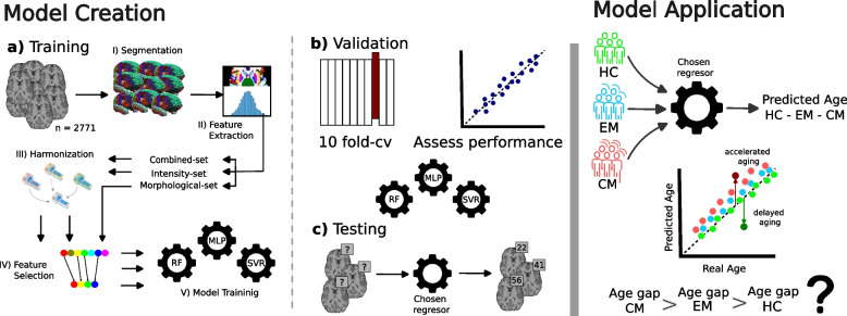
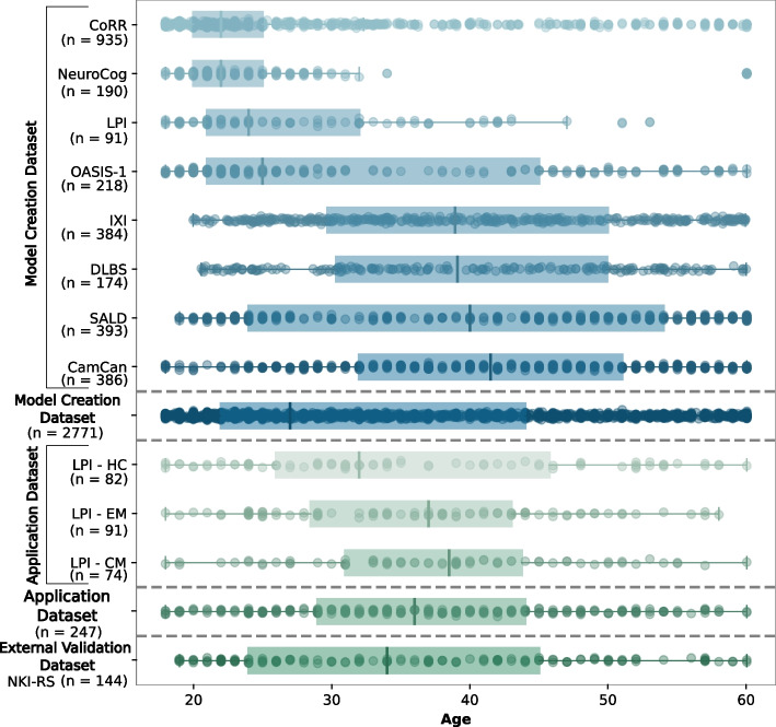
We trained a machine learning model to predict Brain Age from 2,771 T1-weighted magnetic resonance imaging scans of healthy subjects. The processing pipeline included the automatic segmentation of the images, the extraction of 1,479 imaging features (both morphological and intensity-based), harmonization, feature selection and training inside a 10-fold cross-validation scheme. Separate models based only on morphological and intensity features were also trained, and all the Brain Age models were later applied to a discovery cohort composed of 247 subjects, divided into healthy controls (HC, n=82), episodic migraine (EM, n=91), and chronic migraine patients (CM, n=74).
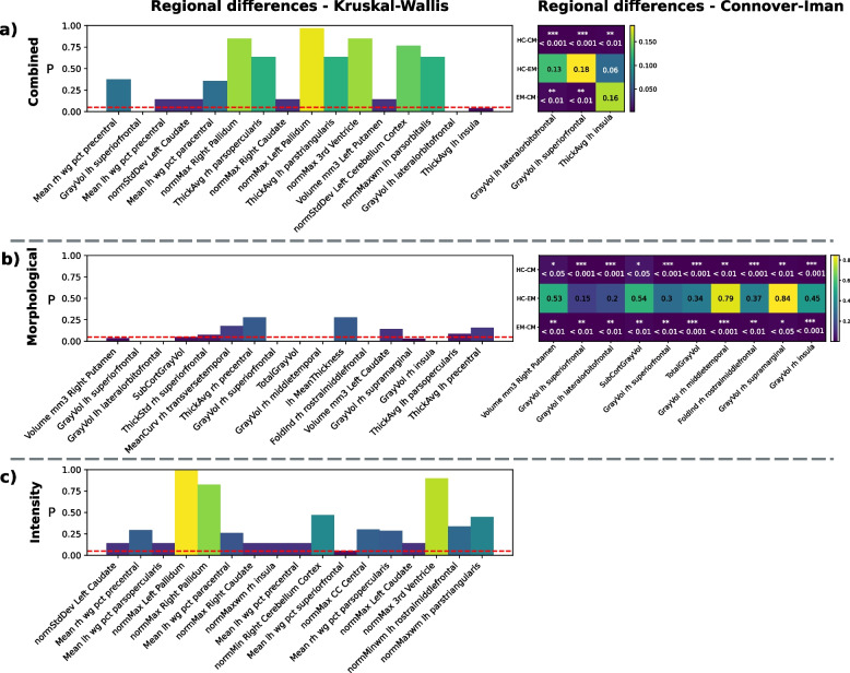
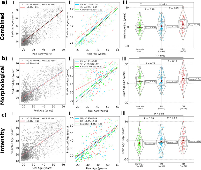
CM patients showed an increased Brain Age Gap compared to HC (4.16 vs -0.56 years, P=0.01). A smaller Brain Age Gap was found for EM patients, not reaching statistical significance (1.21 vs -0.56 years, P=0.19). No associations were found between the Brain Age Gap and headache or migraine frequency, or duration of the disease. Brain imaging features that have previously been associated with migraine were among the main drivers of the differences in the predicted age. Also, the separate analysis using only morphological or intensity-based features revealed different patterns in the Brain Age biomarker in patients with migraine.
The brain-predicted age has shown to be a sensitive biomarker of CM patients and can help reveal distinct aging patterns in migraine.
神经影像学研究表明,偏头痛与大脑结构和功能的改变有关。然而,这些变化与年龄的关系尚未得到详细研究。在这里,我们利用大脑年龄框架来分析偏头痛,构建一个从神经影像学数据预测年龄的机器学习模型。我们假设偏头痛患者的大脑年龄差距(预测年龄与实际年龄的差异)会比健康参与者更大。
我们训练了一个机器学习模型,从 2771 名健康受试者的 T1 加权磁共振成像扫描中预测大脑年龄。处理流水线包括图像的自动分割、1479 个成像特征(形态和强度特征)的提取、调和、特征选择和 10 折交叉验证方案内的训练。还单独训练了仅基于形态和强度特征的模型,并且所有的大脑年龄模型都应用于由 247 名受试者组成的发现队列,分为健康对照组(HC,n=82)、发作性偏头痛(EM,n=91)和慢性偏头痛患者(CM,n=74)。
CM 患者的大脑年龄差距比 HC 患者大(4.16 比-0.56 岁,P=0.01)。EM 患者的大脑年龄差距较小,但没有达到统计学意义(1.21 比-0.56 岁,P=0.19)。大脑年龄差距与头痛或偏头痛频率或疾病持续时间之间没有关联。以前与偏头痛相关的大脑成像特征是预测年龄差异的主要驱动因素之一。此外,仅使用形态或强度特征进行的单独分析显示出偏头痛患者大脑年龄生物标志物的不同模式。
大脑预测年龄已被证明是 CM 患者的敏感生物标志物,可以帮助揭示偏头痛的不同衰老模式。