• 文献检索
  • 文档翻译
  • 深度研究
  • 学术资讯
  • Suppr Zotero 插件Zotero 插件
  • 邀请有礼
  • 套餐&价格
  • 历史记录
应用&插件
Suppr Zotero 插件Zotero 插件浏览器插件Mac 客户端Windows 客户端微信小程序
定价
高级版会员购买积分包购买API积分包
服务
文献检索文档翻译深度研究API 文档MCP 服务
关于我们
关于 Suppr公司介绍联系我们用户协议隐私条款
关注我们

Suppr 超能文献

核心技术专利:CN118964589B侵权必究
粤ICP备2023148730 号-1Suppr @ 2026

文献检索

告别复杂PubMed语法,用中文像聊天一样搜索,搜遍4000万医学文献。AI智能推荐,让科研检索更轻松。

立即免费搜索

文件翻译

保留排版,准确专业,支持PDF/Word/PPT等文件格式,支持 12+语言互译。

免费翻译文档

深度研究

AI帮你快速写综述,25分钟生成高质量综述,智能提取关键信息,辅助科研写作。

立即免费体验

使用基于 CT 的放射组学和深度学习对胃肠道肝转移瘤进行分类。

Tumor classification of gastrointestinal liver metastases using CT-based radiomics and deep learning.

机构信息

Department of Radiology and Nuclear Medicine, University Medical Center Mannheim, Heidelberg University, Theodor-Kutzer-Ufer 1-3, 68167, Mannheim, Germany.

German Cancer Research Center, E010 Radiology, Im Neuenheimer Feld 280, 69120, Heidelberg, Germany.

出版信息

Cancer Imaging. 2023 Oct 5;23(1):95. doi: 10.1186/s40644-023-00612-4.

DOI:10.1186/s40644-023-00612-4
PMID:37798797
原文链接:https://pmc.ncbi.nlm.nih.gov/articles/PMC10557291/
Abstract

OBJECTIVES

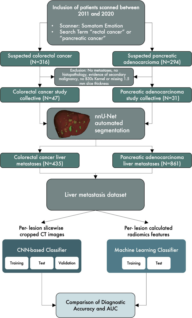
The goal of this study is to demonstrate the performance of radiomics and CNN-based classifiers in determining the primary origin of gastrointestinal liver metastases for visually indistinguishable lesions.

METHODS

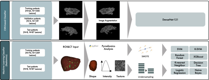
In this retrospective, IRB-approved study, 31 pancreatic cancer patients with 861 lesions (median age [IQR]: 65.39 [56.87, 75.08], 48.4% male) and 47 colorectal cancer patients with 435 lesions (median age [IQR]: 65.79 [56.99, 74.62], 63.8% male) were enrolled. A pretrained nnU-Net performed automated segmentation of 1296 liver lesions. Radiomics features for each lesion were extracted using pyradiomics. The performance of several radiomics-based machine-learning classifiers was investigated for the lesions and compared to an image-based deep-learning approach using a DenseNet-121. The performance was evaluated by AUC/ROC analysis.

RESULTS

The radiomics-based K-nearest neighbor classifier showed the best performance on an independent test set with AUC values of 0.87 and an accuracy of 0.67. In comparison, the image-based DenseNet-121-classifier reached an AUC of 0.80 and an accuracy of 0.83.

CONCLUSIONS

CT-based radiomics and deep learning can distinguish the etiology of liver metastases from gastrointestinal primary tumors. Compared to deep learning, radiomics based models showed a varying generalizability in distinguishing liver metastases from colorectal cancer and pancreatic adenocarcinoma.

摘要

目的

本研究旨在展示基于放射组学和 CNN 的分类器在确定视觉上无法区分的胃肠道肝转移病灶的原发来源方面的性能。

方法

在这项回顾性的、经 IRB 批准的研究中,纳入了 31 名胰腺癌患者的 861 个病灶(中位年龄 [IQR]:65.39 [56.87,75.08],48.4%为男性)和 47 名结直肠癌患者的 435 个病灶(中位年龄 [IQR]:65.79 [56.99,74.62],63.8%为男性)。预训练的 nnU-Net 对 1296 个肝脏病变进行了自动分割。使用 pyradiomics 提取每个病变的放射组学特征。研究了几种基于放射组学的机器学习分类器在病变中的性能,并与基于 DenseNet-121 的图像深度学习方法进行了比较。通过 AUC/ROC 分析评估了性能。

结果

基于放射组学的 K-最近邻分类器在独立测试集上表现最佳,AUC 值为 0.87,准确率为 0.67。相比之下,基于图像的 DenseNet-121 分类器达到了 0.80 的 AUC 和 0.83 的准确率。

结论

基于 CT 的放射组学和深度学习可以区分胃肠道原发肿瘤和肝转移瘤的病因。与深度学习相比,基于放射组学的模型在区分结直肠癌和胰腺腺癌肝转移瘤方面表现出不同的泛化能力。

https://cdn.ncbi.nlm.nih.gov/pmc/blobs/e157/10557291/76d9aeac2257/40644_2023_612_Figa_HTML.jpg
https://cdn.ncbi.nlm.nih.gov/pmc/blobs/e157/10557291/32305f1a18b1/40644_2023_612_Fig1_HTML.jpg
https://cdn.ncbi.nlm.nih.gov/pmc/blobs/e157/10557291/fdbea0acc7f9/40644_2023_612_Fig3_HTML.jpg
https://cdn.ncbi.nlm.nih.gov/pmc/blobs/e157/10557291/427129aeda28/40644_2023_612_Fig2_HTML.jpg
https://cdn.ncbi.nlm.nih.gov/pmc/blobs/e157/10557291/76d9aeac2257/40644_2023_612_Figa_HTML.jpg

相似文献

1
Tumor classification of gastrointestinal liver metastases using CT-based radiomics and deep learning.使用基于 CT 的放射组学和深度学习对胃肠道肝转移瘤进行分类。
Cancer Imaging. 2023 Oct 5;23(1):95. doi: 10.1186/s40644-023-00612-4.
2
Machine learning and radiomics analysis by computed tomography in colorectal liver metastases patients for RAS mutational status prediction.基于 CT 的机器学习和放射组学分析预测结直肠癌肝转移患者的 RAS 基因突变状态。
Radiol Med. 2024 Jul;129(7):957-966. doi: 10.1007/s11547-024-01828-5. Epub 2024 May 18.
3
Deep Learning Radiomics Model of Contrast-Enhanced CT for Differentiating the Primary Source of Liver Metastases.深度学习增强 CT 影像组学模型鉴别肝脏转移瘤的原发灶来源
Acad Radiol. 2024 Oct;31(10):4057-4067. doi: 10.1016/j.acra.2024.04.012. Epub 2024 May 3.
4
CT-Based Radiomics Analysis Before Thermal Ablation to Predict Local Tumor Progression for Colorectal Liver Metastases.基于 CT 的放射组学分析在热消融前预测结直肠癌肝转移的局部肿瘤进展。
Cardiovasc Intervent Radiol. 2021 Jun;44(6):913-920. doi: 10.1007/s00270-020-02735-8. Epub 2021 Jan 27.
5
Distinguishing pure histopathological growth patterns of colorectal liver metastases on CT using deep learning and radiomics: a pilot study.使用深度学习和放射组学在 CT 上区分结直肠癌肝转移的纯组织病理学生长模式:一项初步研究。
Clin Exp Metastasis. 2021 Oct;38(5):483-494. doi: 10.1007/s10585-021-10119-6. Epub 2021 Sep 17.
6
Preoperative classification of primary and metastatic liver cancer via machine learning-based ultrasound radiomics.基于机器学习的超声放射组学对原发性和转移性肝癌的术前分类。
Eur Radiol. 2021 Jul;31(7):4576-4586. doi: 10.1007/s00330-020-07562-6. Epub 2021 Jan 14.
7
Machine Learning and Radiomics Analysis for Tumor Budding Prediction in Colorectal Liver Metastases Magnetic Resonance Imaging Assessment.用于结直肠癌肝转移磁共振成像评估中肿瘤芽生预测的机器学习与影像组学分析
Diagnostics (Basel). 2024 Jan 9;14(2):152. doi: 10.3390/diagnostics14020152.
8
Machine learning-based analysis of CT radiomics model for prediction of colorectal metachronous liver metastases.基于机器学习的 CT 影像组学模型分析用于预测结直肠异时性肝转移。
Abdom Radiol (NY). 2021 Jan;46(1):249-256. doi: 10.1007/s00261-020-02624-1.
9
Machine learning-based radiomics analysis of preoperative functional liver reserve with MRI and CT image.基于机器学习的 MRI 和 CT 图像术前功能性肝储备的放射组学分析。
BMC Med Imaging. 2023 Jul 17;23(1):94. doi: 10.1186/s12880-023-01050-1.
10
Deep learning-based radiomics predicts response to chemotherapy in colorectal liver metastases.基于深度学习的放射组学预测结直肠癌肝转移化疗反应。
Med Phys. 2021 Jan;48(1):513-522. doi: 10.1002/mp.14563. Epub 2020 Nov 30.

引用本文的文献

1
Radiomics Signature of Aging Myocardium in Cardiac Photon-Counting Computed Tomography.心脏光子计数计算机断层扫描中衰老心肌的放射组学特征
Diagnostics (Basel). 2025 Jul 16;15(14):1796. doi: 10.3390/diagnostics15141796.
2
Fully automated 3D multi-modal deep learning model for preoperative T-stage prediction of colorectal cancer using F-FDG PET/CT.用于使用F-FDG PET/CT进行结直肠癌术前T分期预测的全自动3D多模态深度学习模型。
Eur J Nucl Med Mol Imaging. 2025 Jul 28. doi: 10.1007/s00259-025-07450-5.
3
An optimized deep learning model based on transperineal ultrasound images for precision diagnosis of female stress urinary incontinence.

本文引用的文献

1
A Comparison of Techniques for Class Imbalance in Deep Learning Classification of Breast Cancer.深度学习乳腺癌分类中类别不平衡技术的比较
Diagnostics (Basel). 2022 Dec 26;13(1):67. doi: 10.3390/diagnostics13010067.
2
Identification of CT Imaging Phenotypes of Colorectal Liver Metastases from Radiomics Signatures-Towards Assessment of Interlesional Tumor Heterogeneity.基于影像组学特征识别结直肠癌肝转移的CT影像表型——用于评估瘤内肿瘤异质性
Cancers (Basel). 2022 Mar 24;14(7):1646. doi: 10.3390/cancers14071646.
3
Radiomics Features of the Spleen as Surrogates for CT-Based Lymphoma Diagnosis and Subtype Differentiation.
一种基于经会阴超声图像的优化深度学习模型,用于女性压力性尿失禁的精准诊断。
Front Med (Lausanne). 2025 Apr 28;12:1564446. doi: 10.3389/fmed.2025.1564446. eCollection 2025.
4
Radiomics-driven spectral profiling of six kidney stone types with monoenergetic CT reconstructions in photon-counting CT.基于能谱CT单能量重建的放射组学驱动的六种肾结石类型光谱分析
Eur Radiol. 2025 Jun;35(6):3120-3130. doi: 10.1007/s00330-024-11262-w. Epub 2024 Dec 12.
5
Textural heterogeneity of liver lesions in CT imaging - comparison of colorectal and pancreatic metastases.肝脏病变的 CT 影像学纹理异质性——结直肠与胰腺转移瘤的比较。
Abdom Radiol (NY). 2024 Dec;49(12):4295-4306. doi: 10.1007/s00261-024-04511-5. Epub 2024 Aug 8.
6
Differentiation of granulomatous nodules with lobulation and spiculation signs from solid lung adenocarcinomas using a CT deep learning model.利用 CT 深度学习模型鉴别具有分叶和棘突征的肉芽肿性结节与实性肺腺癌。
BMC Cancer. 2024 Jul 22;24(1):875. doi: 10.1186/s12885-024-12611-0.
7
Machine Learning and Radiomics Analysis for Tumor Budding Prediction in Colorectal Liver Metastases Magnetic Resonance Imaging Assessment.用于结直肠癌肝转移磁共振成像评估中肿瘤芽生预测的机器学习与影像组学分析
Diagnostics (Basel). 2024 Jan 9;14(2):152. doi: 10.3390/diagnostics14020152.
脾脏的影像组学特征作为基于CT的淋巴瘤诊断及亚型鉴别替代指标
Cancers (Basel). 2022 Jan 29;14(3):713. doi: 10.3390/cancers14030713.
4
Radiomics and Radiogenomics in Evaluation of Colorectal Cancer Liver Metastasis.放射组学和放射基因组学在结直肠癌肝转移评估中的应用
Front Oncol. 2022 Jan 7;11:689509. doi: 10.3389/fonc.2021.689509. eCollection 2021.
5
Quantitative Imaging Biomarkers of the Whole Liver Tumor Burden Improve Survival Prediction in Metastatic Pancreatic Cancer.全肝肿瘤负荷的定量成像生物标志物可改善转移性胰腺癌的生存预测。
Cancers (Basel). 2021 Nov 16;13(22):5732. doi: 10.3390/cancers13225732.
6
Virtual Biopsy for Diagnosis of Chemotherapy-Associated Liver Injuries and Steatohepatitis: A Combined Radiomic and Clinical Model in Patients with Colorectal Liver Metastases.用于诊断化疗相关肝损伤和脂肪性肝炎的虚拟活检:结直肠癌肝转移患者的放射组学与临床联合模型
Cancers (Basel). 2021 Jun 20;13(12):3077. doi: 10.3390/cancers13123077.
7
Radiogenomic Analysis of Locally Advanced Lung Cancer Based on CT Imaging and Intratreatment Changes in Cell-Free DNA.基于 CT 成像和治疗中游离 DNA 变化的局部晚期肺癌的放射基因组分析。
Radiol Imaging Cancer. 2021 Apr;3(4):e200157. doi: 10.1148/rycan.2021200157.
8
Combining radiomic phenotypes of non-small cell lung cancer with liquid biopsy data may improve prediction of response to EGFR inhibitors.将非小细胞肺癌的放射组学表型与液体活检数据相结合,可能提高对表皮生长因子受体抑制剂反应的预测。
Sci Rep. 2021 May 11;11(1):9984. doi: 10.1038/s41598-021-88239-y.
9
Liver metastases.肝转移。
Nat Rev Dis Primers. 2021 Apr 15;7(1):27. doi: 10.1038/s41572-021-00261-6.
10
Automatic Detection and Classification of Focal Liver Lesions Based on Deep Convolutional Neural Networks: A Preliminary Study.基于深度卷积神经网络的肝脏局灶性病变自动检测与分类:一项初步研究。
Front Oncol. 2021 Jan 29;10:581210. doi: 10.3389/fonc.2020.581210. eCollection 2020.