Hertel Alexander, Froelich Matthias F, Overhoff Daniel, Nestler Tim, Faby Sebastian, Jürgens Markus, Schmidt Bernhard, Vellala Abhinay, Hesse Albrecht, Nörenberg Dominik, Stoll Rico, Schmelz Hans, Schoenberg Stefan O, Waldeck Stephan
Department of Radiology and Nuclear Medicine, University Medical Center Mannheim, University of Heidelberg, Mannheim, Germany.
Department of Diagnostic and Interventional Radiology, Federal Armed Services Hospital Koblenz, Koblenz, Germany.
Eur Radiol. 2025 Jun;35(6):3120-3130. doi: 10.1007/s00330-024-11262-w. Epub 2024 Dec 12.
Urolithiasis, a common and painful urological condition, is influenced by factors such as lifestyle, genetics, and medication. Differentiating between different types of kidney stones is crucial for personalized therapy. The purpose of this study is to investigate the use of photon-counting computed tomography (PCCT) in combination with radiomics and machine learning to develop a method for automated and detailed characterization of kidney stones. This approach aims to enhance the accuracy and detail of stone classification beyond what is achievable with conventional computed tomography (CT) and dual-energy CT (DECT).
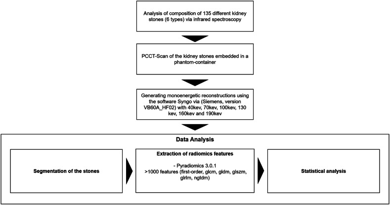
In this ex vivo study, 135 kidney stones were first classified using infrared spectroscopy. All stones were then scanned in a PCCT embedded in a phantom. Various monoenergetic reconstructions were generated, and radiomics features were extracted. Statistical analysis was performed using Random Forest (RF) classifiers for both individual reconstructions and a combined model.
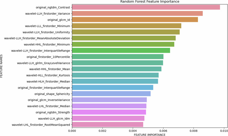
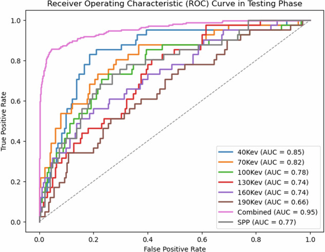
The combined model, using radiomics features from all monoenergetic reconstructions, significantly outperformed individual reconstructions and SPP parameters, with an AUC of 0.95 and test accuracy of 0.81 for differentiating all six stone types. Feature importance analysis identified key parameters, including NGTDM_Strength and wavelet-LLH_firstorder_Variance.
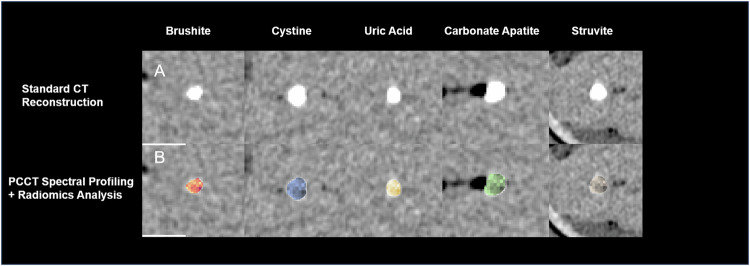
This ex vivo study demonstrates that radiomics-driven PCCT analysis can improve differentiation between kidney stone subtypes. The combined model outperformed individual monoenergetic levels, highlighting the potential of spectral profiling in PCCT to optimize treatment through image-based strategies.
Question How can photon-counting computed tomography (PCCT) combined with radiomics improve the differentiation of kidney stone types beyond conventional CT and dual-energy CT, enhancing personalized therapy? Findings Our ex vivo study demonstrates that a combined spectral-driven radiomics model achieved 95% AUC and 81% test accuracy in differentiating six kidney stone types. Clinical relevance Implementing PCCT-based spectral-driven radiomics allows for precise non-invasive differentiation of kidney stone types, leading to improved diagnostic accuracy and more personalized, effective treatment strategies, potentially reducing the need for invasive procedures and recurrence.
尿石症是一种常见且疼痛的泌尿系统疾病,受生活方式、遗传和药物等因素影响。区分不同类型的肾结石对于个性化治疗至关重要。本研究的目的是探讨光子计数计算机断层扫描(PCCT)结合放射组学和机器学习,以开发一种对肾结石进行自动和详细特征描述的方法。这种方法旨在提高结石分类的准确性和细节程度,超越传统计算机断层扫描(CT)和双能CT(DECT)所能达到的水平。
在这项离体研究中,首先使用红外光谱对135颗肾结石进行分类。然后将所有结石在嵌入体模的PCCT中进行扫描。生成各种单能重建图像,并提取放射组学特征。使用随机森林(RF)分类器对单个重建图像和组合模型进行统计分析。
使用来自所有单能重建图像的放射组学特征的组合模型,在区分所有六种结石类型方面显著优于单个重建图像和SPP参数,曲线下面积(AUC)为0.95,测试准确率为0.81。特征重要性分析确定了关键参数,包括NGTDM_Strength和小波-LLH_firstorder_Variance。
这项离体研究表明,放射组学驱动的PCCT分析可改善肾结石亚型之间的区分。组合模型优于单个单能水平,突出了PCCT中光谱分析通过基于图像的策略优化治疗的潜力。
问题 光子计数计算机断层扫描(PCCT)结合放射组学如何超越传统CT和双能CT提高肾结石类型的区分能力,增强个性化治疗?研究结果我们的离体研究表明,光谱驱动的放射组学组合模型在区分六种肾结石类型时AUC达到95%,测试准确率达到81%。临床意义实施基于PCCT的光谱驱动放射组学可实现肾结石类型的精确无创区分,提高诊断准确性,制定更个性化、有效的治疗策略,可能减少侵入性手术的需求和复发率。