• 文献检索
  • 文档翻译
  • 深度研究
  • 学术资讯
  • Suppr Zotero 插件Zotero 插件
  • 邀请有礼
  • 套餐&价格
  • 历史记录
应用&插件
Suppr Zotero 插件Zotero 插件浏览器插件Mac 客户端Windows 客户端微信小程序
定价
高级版会员购买积分包购买API积分包
服务
文献检索文档翻译深度研究API 文档MCP 服务
关于我们
关于 Suppr公司介绍联系我们用户协议隐私条款
关注我们

Suppr 超能文献

核心技术专利:CN118964589B侵权必究
粤ICP备2023148730 号-1Suppr @ 2026

文献检索

告别复杂PubMed语法,用中文像聊天一样搜索,搜遍4000万医学文献。AI智能推荐,让科研检索更轻松。

立即免费搜索

文件翻译

保留排版,准确专业,支持PDF/Word/PPT等文件格式,支持 12+语言互译。

免费翻译文档

深度研究

AI帮你快速写综述,25分钟生成高质量综述,智能提取关键信息,辅助科研写作。

立即免费体验

通过 Glycophorin C 定量和可视化的红细胞积累反映了斑块内出血和术前神经症状。

The accumulation of erythrocytes quantified and visualized by Glycophorin C in carotid atherosclerotic plaque reflects intraplaque hemorrhage and pre-procedural neurological symptoms.

机构信息

Division of Surgical Specialties, Department of Vascular Surgery, University Medical Center Utrecht, Utrecht University, Heidelberglaan 100, 3508 GA, Utrecht, The Netherlands.

Laboratory of Experimental Cardiology, Department of Cardiology, Division Heart and Lungs, University Medical Center Utrecht, Utrecht University, Heidelberglaan 100, 3508 GA, Utrecht, The Netherlands.

出版信息

Sci Rep. 2023 Oct 10;13(1):17104. doi: 10.1038/s41598-023-43369-3.

DOI:10.1038/s41598-023-43369-3
PMID:37816779
原文链接:https://pmc.ncbi.nlm.nih.gov/articles/PMC10564864/
Abstract

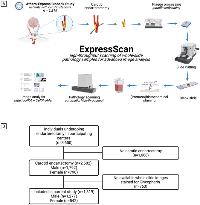
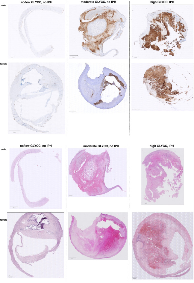
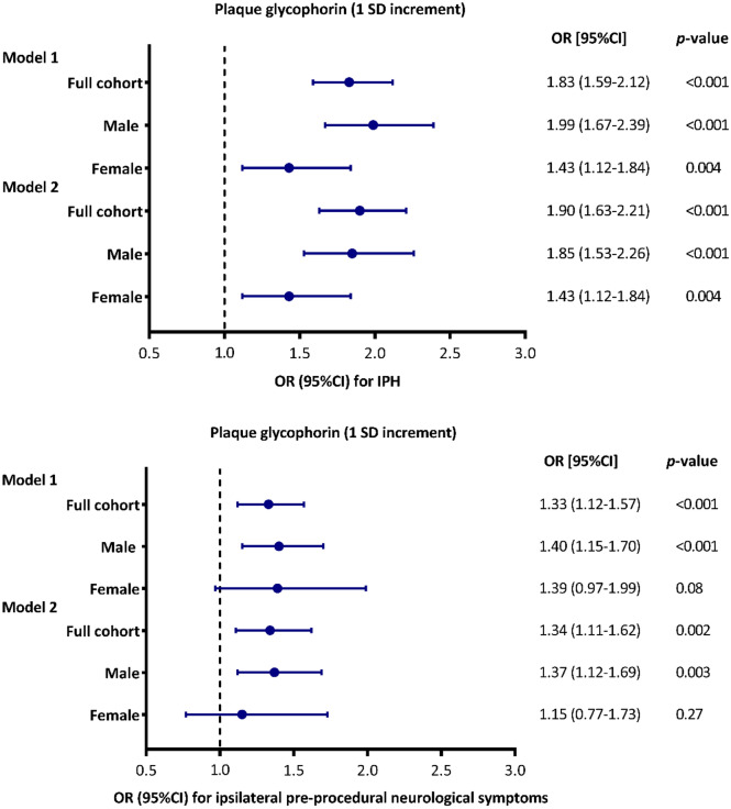
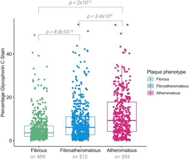
The accumulation of erythrocyte membranes within an atherosclerotic plaque may contribute to the deposition of free cholesterol and thereby the enlargement of the necrotic core. Erythrocyte membranes can be visualized and quantified in the plaque by immunostaining for the erythrocyte marker glycophorin C. Hence, we theorized that the accumulation of erythrocytes quantified by glycophorin C could function as a marker for plaque vulnerability, possibly reflecting intraplaque hemorrhage (IPH), and offering predictive value for pre-procedural neurological symptoms. We employed the CellProfiler-integrated slideToolKit workflow to visualize and quantify glycophorin C, defined as the total plaque area that is positive for glycophorin C, in single slides of culprit lesions obtained from the Athero-Express Biobank of 1819 consecutive asymptomatic and symptomatic patients who underwent carotid endarterectomy. Our assessment included the evaluation of various parameters such as lipid core, calcifications, collagen content, SMC content, and macrophage burden. These parameters were evaluated using a semi-quantitative scoring method, and the resulting data was dichotomized as predefined criteria into categories of no/minor or moderate/heavy staining. In addition, the presence or absence of IPH was also scored. The prevalence of IPH and pre-procedural neurological symptoms were 62.4% and 87.1%, respectively. The amount of glycophorin staining was significantly higher in samples from men compared to samples of women (median 7.15 (IQR:3.37, 13.41) versus median 4.06 (IQR:1.98, 8.32), p < 0.001). Glycophorin C was associated with IPH adjusted for clinical confounders (OR 1.90; 95% CI 1.63, 2.21; p =  < 0.001). Glycophorin C was significantly associated with ipsilateral pre-procedural neurological symptoms (OR:1.27, 95%CI:1.06-1.41, p = 0.005). Sex-stratified analysis, showed that this was also the case for men (OR 1.37; 95%CI 1.12, 1.69; p = 0.003), but not for women (OR 1.15; 95%CI 0.77, 1.73; p = 0.27). Glycophorin C was associated with classical features of a vulnerable plaque, such as a larger lipid core, a higher macrophage burden, less calcifications, a lower collagen and SMC content. There were marked sex differences, in men, glycophorin C was associated with calcifications and collagen while these associations were not found in women. To conclude, the accumulation of erythrocytes in atherosclerotic plaque quantified and visualized by glycophorin C was independently associated with the presence of IPH, preprocedural symptoms in men, and with a more vulnerable plaque composition in both men and women. These results strengthen the notion that the accumulation of erythrocytes quantified by glycophorin C can be used as a marker for plaque vulnerability.

摘要

动脉粥样硬化斑块内红细胞膜的积累可能有助于游离胆固醇的沉积,从而导致坏死核心的增大。通过对红细胞标志物糖蛋白 C 的免疫染色,可以在斑块中可视化和定量红细胞膜。因此,我们推测通过糖蛋白 C 定量的红细胞积累可以作为斑块脆弱性的标志物,可能反映斑块内出血 (IPH),并为术前神经症状提供预测价值。我们使用集成了 CellProfiler 的 slideToolKit 工作流程来可视化和定量糖蛋白 C,糖蛋白 C 定义为糖蛋白 C 阳性的总斑块面积,在从接受颈动脉内膜切除术的 1819 名无症状和有症状的连续患者的动脉粥样硬化表达生物库中获得的罪魁祸首病变的单个幻灯片中进行定量。我们的评估包括评估各种参数,如脂质核心、钙化、胶原蛋白含量、SMC 含量和巨噬细胞负担。这些参数使用半定量评分方法进行评估,并根据预先定义的标准将结果数据分为无/轻度或中度/重度染色的类别。此外,还对 IPH 的存在进行了评分。IPH 和术前神经症状的发生率分别为 62.4%和 87.1%。与女性样本相比,男性样本中的糖蛋白染色量明显更高(中位数 7.15(IQR:3.37,13.41)与中位数 4.06(IQR:1.98,8.32)相比,p<0.001)。在调整了临床混杂因素后,糖蛋白 C 与 IPH 相关(OR 1.90;95%CI 1.63,2.21;p<0.001)。糖蛋白 C 与同侧术前神经症状显著相关(OR:1.27,95%CI:1.06-1.41,p=0.005)。按性别分层分析表明,这对男性也是如此(OR 1.37;95%CI 1.12,1.69;p=0.003),但对女性则不然(OR 1.15;95%CI 0.77,1.73;p=0.27)。糖蛋白 C 与易损斑块的典型特征相关,例如更大的脂质核心、更高的巨噬细胞负担、更少的钙化、更低的胶原蛋白和 SMC 含量。存在明显的性别差异,在男性中,糖蛋白 C 与钙化和胶原蛋白相关,而在女性中则没有发现这些关联。总之,通过糖蛋白 C 定量和可视化的动脉粥样硬化斑块中红细胞的积累与男性中的 IPH、术前症状以及男性和女性中更易损的斑块成分独立相关。这些结果进一步证实了通过糖蛋白 C 定量的红细胞积累可以作为斑块脆弱性的标志物的观点。

https://cdn.ncbi.nlm.nih.gov/pmc/blobs/dd34/10564864/642b2b54d195/41598_2023_43369_Fig7_HTML.jpg
https://cdn.ncbi.nlm.nih.gov/pmc/blobs/dd34/10564864/f6989271e369/41598_2023_43369_Fig1_HTML.jpg
https://cdn.ncbi.nlm.nih.gov/pmc/blobs/dd34/10564864/ab779bc74a46/41598_2023_43369_Fig2_HTML.jpg
https://cdn.ncbi.nlm.nih.gov/pmc/blobs/dd34/10564864/9282a97fa05b/41598_2023_43369_Fig3_HTML.jpg
https://cdn.ncbi.nlm.nih.gov/pmc/blobs/dd34/10564864/b4fd8f12841f/41598_2023_43369_Fig4_HTML.jpg
https://cdn.ncbi.nlm.nih.gov/pmc/blobs/dd34/10564864/4c123f794f33/41598_2023_43369_Fig5_HTML.jpg
https://cdn.ncbi.nlm.nih.gov/pmc/blobs/dd34/10564864/0b52e32b031a/41598_2023_43369_Fig6_HTML.jpg
https://cdn.ncbi.nlm.nih.gov/pmc/blobs/dd34/10564864/642b2b54d195/41598_2023_43369_Fig7_HTML.jpg

相似文献

1
The accumulation of erythrocytes quantified and visualized by Glycophorin C in carotid atherosclerotic plaque reflects intraplaque hemorrhage and pre-procedural neurological symptoms.通过 Glycophorin C 定量和可视化的红细胞积累反映了斑块内出血和术前神经症状。
Sci Rep. 2023 Oct 10;13(1):17104. doi: 10.1038/s41598-023-43369-3.
2
Sex Differences in Plaque Composition and Morphology Among Symptomatic Patients With Mild-to-Moderate Carotid Artery Stenosis.症状性轻至中度颈动脉狭窄患者斑块成分和形态的性别差异。
Stroke. 2022 Feb;53(2):370-378. doi: 10.1161/STROKEAHA.121.036564. Epub 2022 Jan 5.
3
Magnetic Resonance Imaging Identified Brain Ischaemia in Symptomatic Patients Undergoing Carotid Endarterectomy Is Related to Histologically Apparent Intraplaque Haemorrhage.磁共振成像在接受颈动脉内膜切除术的有症状患者中识别到与组织学上明显的斑块内出血有关的脑缺血。
Eur J Vasc Endovasc Surg. 2019 Dec;58(6):796-804. doi: 10.1016/j.ejvs.2019.07.017. Epub 2019 Oct 18.
4
Superficial and multiple calcifications and ulceration associate with intraplaque hemorrhage in the carotid atherosclerotic plaque.颈动脉粥样硬化斑块内斑块出血与表面及多发钙化和溃疡相关。
Eur Radiol. 2018 Dec;28(12):4968-4977. doi: 10.1007/s00330-018-5535-7. Epub 2018 Jun 6.
5
Different stages of intraplaque hemorrhage are associated with different plaque phenotypes: a large histopathological study in 794 carotid and 276 femoral endarterectomy specimens.不同阶段的斑块内出血与不同的斑块表型相关:794 例颈动脉和 276 例股动脉内膜切除术标本的大型组织病理学研究。
Atherosclerosis. 2011 Oct;218(2):369-77. doi: 10.1016/j.atherosclerosis.2011.07.104. Epub 2011 Jul 30.
6
Preoperative hypertension is associated with atherosclerotic intraplaque hemorrhage in patients undergoing carotid endarterectomy.术前高血压与颈动脉内膜切除术患者的粥样硬化斑块内出血有关。
Atherosclerosis. 2019 Nov;290:214-221. doi: 10.1016/j.atherosclerosis.2019.09.008. Epub 2019 Sep 22.
7
Ipsilateral plaques display higher T1 signals than contralateral plaques in recently symptomatic patients with bilateral carotid intraplaque hemorrhage.在近期出现症状的双侧颈动脉斑块内出血患者中,同侧斑块的T1信号高于对侧斑块。
Atherosclerosis. 2017 Feb;257:78-85. doi: 10.1016/j.atherosclerosis.2017.01.001. Epub 2017 Jan 12.
8
Assessment of necrotic core with intraplaque hemorrhage in atherosclerotic carotid artery plaque by MR imaging with 3D gradient-echo sequence in patients with high-grade stenosis. Clinical article.高狭窄程度患者应用 3D 梯度回波序列磁共振成像评估动脉粥样硬化性颈动脉斑块内的坏死核心伴斑块内出血。临床文章。
J Neurosurg. 2010 Oct;113(4):890-6. doi: 10.3171/2010.3.JNS091057.
9
Quantitative evaluation of high intensity signal on MIP images of carotid atherosclerotic plaques from routine TOF-MRA reveals elevated volumes of intraplaque hemorrhage and lipid rich necrotic core.颈动脉粥样硬化斑块 MIP 图像高强度信号的定量评估显示,斑块内出血和富含脂质的坏死核心体积增加。
J Cardiovasc Magn Reson. 2012 Nov 29;14(1):81. doi: 10.1186/1532-429X-14-81.
10
Determinants of carotid atherosclerotic plaque burden in a stroke-free population.无卒中人群中颈动脉粥样硬化斑块负荷的决定因素。
Atherosclerosis. 2016 Dec;255:186-192. doi: 10.1016/j.atherosclerosis.2016.10.030. Epub 2016 Oct 14.

引用本文的文献

1
Smooth muscle expression of RNA editing enzyme ADAR1 controls activation of the RNA sensor MDA5 in atherosclerosis.RNA编辑酶ADAR1的平滑肌表达调控动脉粥样硬化中RNA传感器MDA5的激活。
Nat Cardiovasc Res. 2025 Sep 16. doi: 10.1038/s44161-025-00710-5.
2
Intraplaque haemorrhage quantification and molecular characterisation using attention based multiple instance learning.基于注意力的多实例学习用于斑块内出血定量分析和分子特征描述
medRxiv. 2025 Mar 26:2025.03.04.25323316. doi: 10.1101/2025.03.04.25323316.
3
The Role of Red Blood Cells in Cholesterol Accumulation and Atherosclerotic Plaque Instability: A Perspective on Atherosclerosis.

本文引用的文献

1
Editor's Choice - European Society for Vascular Surgery (ESVS) 2023 Clinical Practice Guidelines on the Management of Atherosclerotic Carotid and Vertebral Artery Disease.编辑推荐——欧洲血管外科学会(ESVS)2023年动脉粥样硬化性颈动脉和椎动脉疾病管理临床实践指南
Eur J Vasc Endovasc Surg. 2023 Jan;65(1):7-111. doi: 10.1016/j.ejvs.2022.04.011. Epub 2022 May 20.
2
False Utopia of One Unifying Description of the Vulnerable Atherosclerotic Plaque: A Call for Recalibration That Appreciates the Diversity of Mechanisms Leading to Atherosclerotic Disease.虚假的脆弱粥样硬化斑块单一描述一统论的乌托邦:呼吁重新校准,以认识到导致动脉粥样硬化疾病的多种机制。
Arterioscler Thromb Vasc Biol. 2022 Apr;42(4):e86-e95. doi: 10.1161/ATVBAHA.121.316693. Epub 2022 Feb 10.
3
红细胞在胆固醇积累和动脉粥样硬化斑块不稳定中的作用:关于动脉粥样硬化的观点
Curr Cardiol Rev. 2025;21(5):22-28. doi: 10.2174/011573403X358572250128104335.
Erythrocytes: Central Actors in Multiple Scenes of Atherosclerosis.红细胞:动脉粥样硬化多幕剧中的主角
Int J Mol Sci. 2021 May 29;22(11):5843. doi: 10.3390/ijms22115843.
4
The age- and sex-specific composition of atherosclerotic plaques in vascular surgery patients.血管外科患者粥样斑块的年龄和性别特异性构成。
Atherosclerosis. 2020 Oct;310:1-10. doi: 10.1016/j.atherosclerosis.2020.07.016. Epub 2020 Jul 29.
5
Pathophysiology of atherothrombosis: Mechanisms of thrombus formation on disrupted atherosclerotic plaques.动脉粥样硬化血栓形成的病理生理学:破裂粥样硬化斑块上血栓形成的机制。
Pathol Int. 2020 Jun;70(6):309-322. doi: 10.1111/pin.12921. Epub 2020 Mar 13.
6
A Farewell to Classic Qualitative Microscope Histology Assessment.告别经典定性显微镜组织学评估。
Eur J Vasc Endovasc Surg. 2020 Mar;59(3):488. doi: 10.1016/j.ejvs.2019.09.504. Epub 2019 Oct 7.
7
Atherosclerosis.动脉粥样硬化。
Nat Rev Dis Primers. 2019 Aug 16;5(1):56. doi: 10.1038/s41572-019-0106-z.
8
Iron and Atherosclerosis: The Link Revisited.铁与动脉粥样硬化:重新审视二者的关联。
Trends Mol Med. 2019 Aug;25(8):659-661. doi: 10.1016/j.molmed.2019.05.012. Epub 2019 Jun 20.
9
Lysed Erythrocyte Membranes Promote Vascular Calcification.溶血性红细胞膜促进血管钙化。
Circulation. 2019 Apr 23;139(17):2032-2048. doi: 10.1161/CIRCULATIONAHA.118.037166.
10
A Comprehensive Review of Our Current Understanding of Red Blood Cell (RBC) Glycoproteins.关于我们目前对红细胞(RBC)糖蛋白理解的全面综述。
Membranes (Basel). 2017 Sep 29;7(4):56. doi: 10.3390/membranes7040056.