• 文献检索
  • 文档翻译
  • 深度研究
  • 学术资讯
  • Suppr Zotero 插件Zotero 插件
  • 邀请有礼
  • 套餐&价格
  • 历史记录
应用&插件
Suppr Zotero 插件Zotero 插件浏览器插件Mac 客户端Windows 客户端微信小程序
定价
高级版会员购买积分包购买API积分包
服务
文献检索文档翻译深度研究API 文档MCP 服务
关于我们
关于 Suppr公司介绍联系我们用户协议隐私条款
关注我们

Suppr 超能文献

核心技术专利:CN118964589B侵权必究
粤ICP备2023148730 号-1Suppr @ 2026

文献检索

告别复杂PubMed语法,用中文像聊天一样搜索,搜遍4000万医学文献。AI智能推荐,让科研检索更轻松。

立即免费搜索

文件翻译

保留排版,准确专业,支持PDF/Word/PPT等文件格式,支持 12+语言互译。

免费翻译文档

深度研究

AI帮你快速写综述,25分钟生成高质量综述,智能提取关键信息,辅助科研写作。

立即免费体验

在制备纳米阵列以提高其表面增强拉曼散射(SERS)性能方面,无表面活性剂的Au@Ag与CTAB稳定的星形Au@Ag纳米颗粒之间的差异。

Differences between surfactant-free Au@Ag and CTAB-stabilized Au@Ag star-like nanoparticles in the preparation of nanoarrays to improve their surface-enhanced Raman scattering (SERS) performance.

作者信息

Van Vu Sy, Nguyen Anh-Thu, Cao Tran Anh-Thi, Thi Le Viet-Ha, Lo Tien Nu Hoang, Ho Thi H, Pham Nguyet N T, Park In, Vo Khuong Quoc

机构信息

Faculty of Chemistry, University of Science, Vietnam National University - Ho Chi Minh City 227 Nguyen Van Cu Street, Ward 4, District 5 Ho Chi Minh City 70000 Vietnam

Vietnam National University Ho Chi Minh City Vietnam.

出版信息

Nanoscale Adv. 2023 Sep 4;5(20):5543-5561. doi: 10.1039/d3na00483j. eCollection 2023 Oct 10.

DOI:10.1039/d3na00483j
PMID:37822906
原文链接:https://pmc.ncbi.nlm.nih.gov/articles/PMC10563836/
Abstract

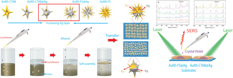
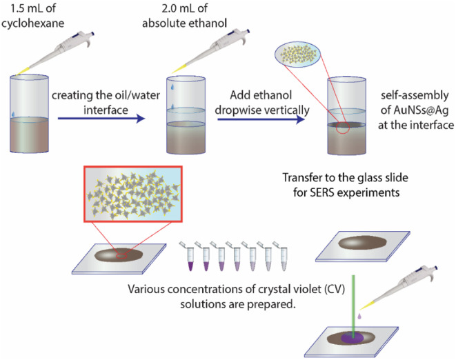
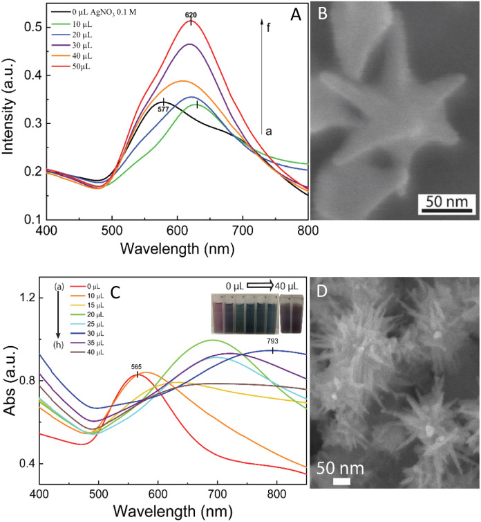
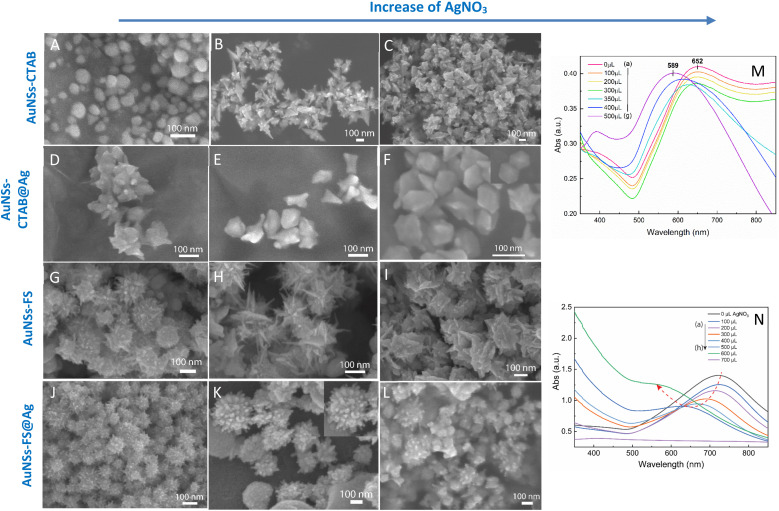
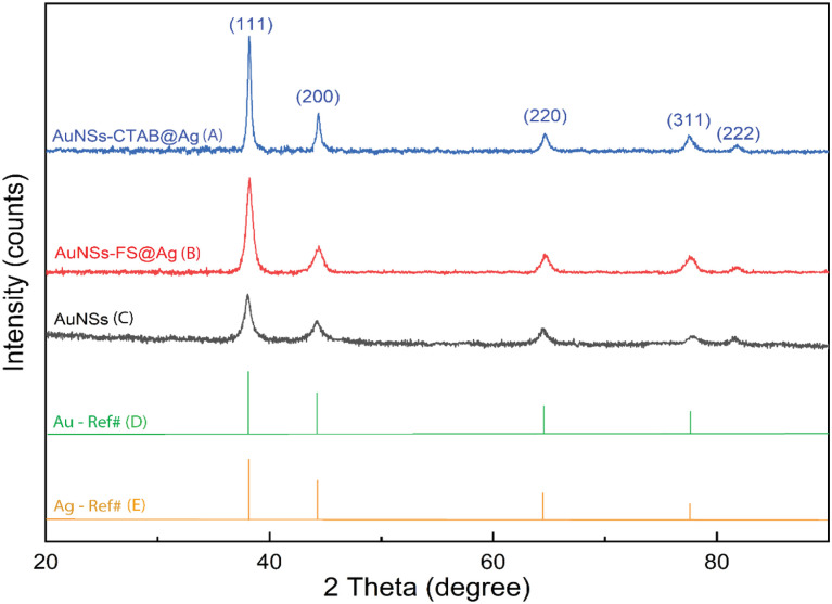
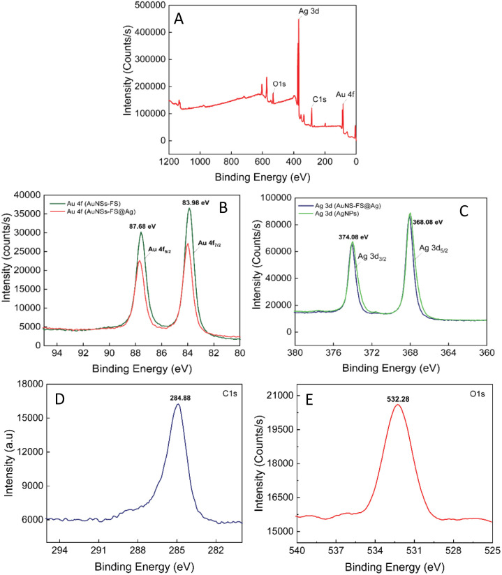
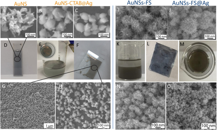
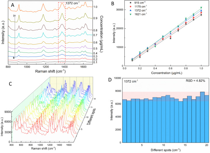
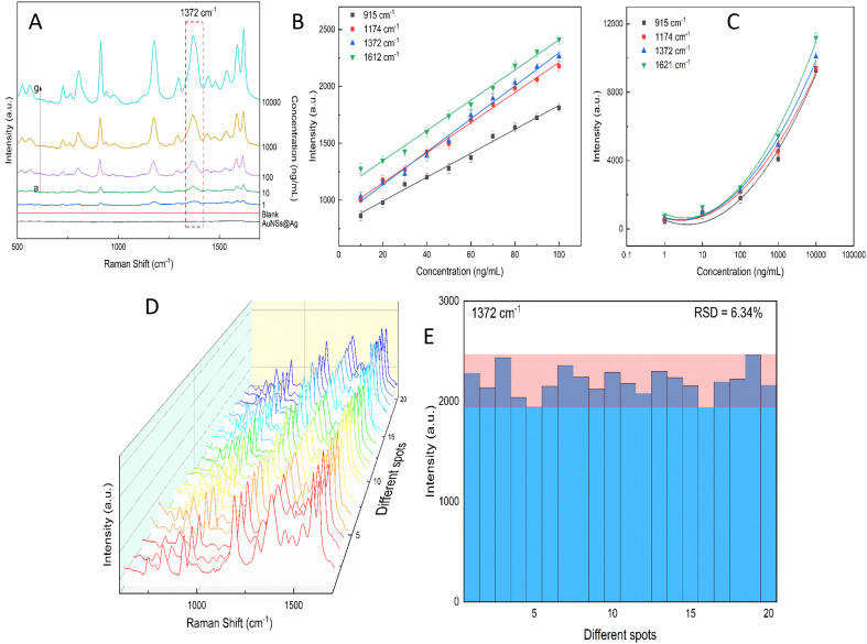
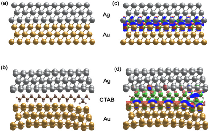
In this study, we assessed the controlled synthesis and efficacy of surface-enhanced Raman scattering (SERS) on two distinct types of star-like Au@Ag core-shell nanoarrays. These nanoarrays were designed based on gold nanostars (AuNSs), which were synthesized with and without CTAB surfactant (AuNSs-CTAB and AuNSs-FS, respectively). The AuNS-FS nanoparticles were synthesized a novel modification process, which helped overcome the previous limitations in the free-surfactant preparation of AuNSs by significantly increasing the number of branches, increasing the sharpness of the branches and minimizing the adsorption of the surfactant on the surface of AuNSs. Furthermore, the differences in the size and morphology of these AuNSs in the created nanoarrays were studied. To create the nanoarrays, a three-step method was employed, which involved the controlled synthesis of gold nanostars, covering them with a silver layer (AuNSs-FS@Ag and AuNSs-CTAB@Ag), and finally self-assembling the AuNS@Ag core-shelled nanoparticles the liquid/liquid self-assembly method. AuNSs-FS@Ag showed higher ability in forming self-assembled nanoarrays than the nanoparticles prepared using CTAB, which can be attributed to the decrease in the repulsion between the nanoparticles at the interface. The nano-substrates developed with AuNSs-FS@Ag possessed numerous "hot spots" on their surface, resulting in a highly effective SERS performance. AuNSs-FS featured a significantly higher number of sharp branches than AuNSs-CTAB, making it the better choice for creating nanoarrays. It is worth mentioning that AuNSs-CTAB did not exhibit the same benefits as AuNSs-FS. The morphology of AuNSs with numerous branches was formed by controlling the seed boiling temperature and adding a specific amount of silver ions. To compare the SERS activity between the as-prepared nano-substrates, , AuNS-CTAB@Ag and AuNS-FS@Ag self-assembled nanoarrays, low concentrations of crystal violet aqueous solution were characterized. The results showed that the developed AuNSs-FS@Ag could detect CV at trace concentrations ranging from 1.0 ng mL to 10 ng mL with a limit of detection (LOD) of 0.45 ng mL and limit of quantification (LOQ) of 1.38 ng mL. The nano-substrates remained stable for 42 days with a decrease in the intensity of the characteristic Raman peaks of CV by less than 7.0% after storage. Furthermore, the spiking method could detect trace amounts of CV in natural water from the Dong Nai River with concentrations as low as 1 to 100 ng mL, with an LOD of 6.07 ng mL and LOQ of 18.4 ng mL. This method also displayed good reproducibility with an RSD value of 5.71%. To better understand the impact of CTAB stabilization of the Au@Ag star-like nanoparticles on their surface-enhanced Raman scattering (SERS) performance, we conducted density functional theory (DFT) calculations. Our research showed that the preparation of AuNSs-FS@Ag self-assembly is an efficient, simple, and fast process, which can be easily performed in any laboratory. Furthermore, the research and development results presented herein on nanoarrays have potential application in analyzing and determining trace amounts of organic compounds in textile dyeing wastewater.

摘要

在本研究中,我们评估了表面增强拉曼散射(SERS)在两种不同类型的星形金@银核壳纳米阵列上的可控合成及效果。这些纳米阵列是基于金纳米星(AuNSs)设计的,分别在有和没有十六烷基三甲基溴化铵(CTAB)表面活性剂的情况下合成(分别为AuNSs - CTAB和AuNSs - FS)。AuNS - FS纳米粒子是通过一种新颖的改性工艺合成的,该工艺通过显著增加分支数量、提高分支的尖锐度并使表面活性剂在AuNSs表面的吸附最小化,帮助克服了先前在无表面活性剂制备AuNSs时的局限性。此外,还研究了这些AuNSs在生成的纳米阵列中的尺寸和形态差异。为了创建纳米阵列,采用了一种三步法,包括金纳米星的可控合成、用银层覆盖它们(AuNSs - FS@Ag和AuNSs - CTAB@Ag),最后通过液/液自组装方法将金@银核壳纳米粒子自组装。与使用CTAB制备的纳米粒子相比,AuNSs - FS@Ag在形成自组装纳米阵列方面表现出更高的能力,这可归因于界面处纳米粒子之间排斥力的降低。用AuNSs - FS@Ag开发的纳米基底表面有许多“热点”,从而产生了高效的SERS性能。AuNSs - FS的尖锐分支数量明显多于AuNSs - CTAB,使其成为创建纳米阵列的更好选择。值得一提的是,AuNSs - CTAB没有表现出与AuNSs - FS相同的优势。通过控制种子沸腾温度并添加特定量的银离子,形成了具有许多分支的AuNSs形态。为了比较所制备的纳米基底(即AuNS - CTAB@Ag和AuNS - FS@Ag自组装纳米阵列)之间的SERS活性,对低浓度的结晶紫水溶液进行了表征。结果表明,所开发的AuNSs - FS@Ag能够检测浓度范围为1.0 ng/mL至10 ng/mL的痕量结晶紫,检测限(LOD)为0.45 ng/mL,定量限(LOQ)为1.38 ng/mL。纳米基底在储存42天后保持稳定,结晶紫特征拉曼峰强度下降小于7.0%。此外,加标法能够检测同奈河天然水中低至1至100 ng/mL的痕量结晶紫,LOD为6.07 ng/mL,LOQ为18.4 ng/mL。该方法还具有良好的重现性,相对标准偏差(RSD)值为5.71%。为了更好地理解CTAB对金@银星形纳米粒子的稳定作用对其表面增强拉曼散射(SERS)性能的影响,我们进行了密度泛函理论(DFT)计算。我们的研究表明,通过自组装制备AuNSs - FS@Ag是一个高效、简单且快速的过程,在任何实验室都能轻松进行。此外,本文介绍的关于纳米阵列的研发成果在分析和测定纺织印染废水中痕量有机化合物方面具有潜在应用。

https://cdn.ncbi.nlm.nih.gov/pmc/blobs/e2d0/10563836/f9ae63a5d218/d3na00483j-f15.jpg
https://cdn.ncbi.nlm.nih.gov/pmc/blobs/e2d0/10563836/0a176b21ef42/d3na00483j-f1.jpg
https://cdn.ncbi.nlm.nih.gov/pmc/blobs/e2d0/10563836/be73305a23a0/d3na00483j-f2.jpg
https://cdn.ncbi.nlm.nih.gov/pmc/blobs/e2d0/10563836/1e37ab33daf5/d3na00483j-f3.jpg
https://cdn.ncbi.nlm.nih.gov/pmc/blobs/e2d0/10563836/a84be2665a10/d3na00483j-f4.jpg
https://cdn.ncbi.nlm.nih.gov/pmc/blobs/e2d0/10563836/d3039266e768/d3na00483j-f5.jpg
https://cdn.ncbi.nlm.nih.gov/pmc/blobs/e2d0/10563836/440cf5a33ea4/d3na00483j-f6.jpg
https://cdn.ncbi.nlm.nih.gov/pmc/blobs/e2d0/10563836/2b72fb68b91c/d3na00483j-f7.jpg
https://cdn.ncbi.nlm.nih.gov/pmc/blobs/e2d0/10563836/871f40d44b7d/d3na00483j-f8.jpg
https://cdn.ncbi.nlm.nih.gov/pmc/blobs/e2d0/10563836/1b4f781daa24/d3na00483j-f9.jpg
https://cdn.ncbi.nlm.nih.gov/pmc/blobs/e2d0/10563836/502af8d20708/d3na00483j-f10.jpg
https://cdn.ncbi.nlm.nih.gov/pmc/blobs/e2d0/10563836/b8c82dc29778/d3na00483j-f11.jpg
https://cdn.ncbi.nlm.nih.gov/pmc/blobs/e2d0/10563836/f7f430c0fe76/d3na00483j-f12.jpg
https://cdn.ncbi.nlm.nih.gov/pmc/blobs/e2d0/10563836/8522b6d2a2fb/d3na00483j-f13.jpg
https://cdn.ncbi.nlm.nih.gov/pmc/blobs/e2d0/10563836/36a49efecfe4/d3na00483j-f14.jpg
https://cdn.ncbi.nlm.nih.gov/pmc/blobs/e2d0/10563836/f9ae63a5d218/d3na00483j-f15.jpg

相似文献

1
Differences between surfactant-free Au@Ag and CTAB-stabilized Au@Ag star-like nanoparticles in the preparation of nanoarrays to improve their surface-enhanced Raman scattering (SERS) performance.在制备纳米阵列以提高其表面增强拉曼散射(SERS)性能方面,无表面活性剂的Au@Ag与CTAB稳定的星形Au@Ag纳米颗粒之间的差异。
Nanoscale Adv. 2023 Sep 4;5(20):5543-5561. doi: 10.1039/d3na00483j. eCollection 2023 Oct 10.
2
Gold nanostars: Benzyldimethylammonium chloride-assisted synthesis, plasmon tuning, SERS and catalytic activity.金纳米星:苄基二甲基氯化铵辅助合成、等离子体调谐、表面增强拉曼光谱及催化活性
J Colloid Interface Sci. 2016 Jan 15;462:341-50. doi: 10.1016/j.jcis.2015.10.007. Epub 2015 Oct 9.
3
Facile tuning of tip sharpness on gold nanostars by the controlled seed-growth method and coating with a silver shell for detection of thiram using surface enhanced Raman spectroscopy (SERS).通过可控种子生长法轻松调节金纳米星的尖端锐度,并包覆银壳以利用表面增强拉曼光谱(SERS)检测福美双。
RSC Adv. 2022 Aug 15;12(35):22815-22825. doi: 10.1039/d2ra03396h. eCollection 2022 Aug 10.
4
Zinc oxide/silver nanoarrays as reusable SERS substrates with controllable 'hot-spots' for highly reproducible molecular sensing.氧化锌/银纳米阵列作为可重复使用的 SERS 基底,具有可控的“热点”,可实现高度重现的分子传感。
J Colloid Interface Sci. 2014 Dec 15;436:251-7. doi: 10.1016/j.jcis.2014.09.017. Epub 2014 Sep 17.
5
High-sensitive bioorthogonal SERS tag for live cancer cell imaging by self-assembling core-satellites structure gold-silver nanocomposite.高灵敏度生物正交 SERS 标签通过自组装核-卫星结构金银纳米复合材料用于活癌细胞成像。
Talanta. 2017 Sep 1;172:176-181. doi: 10.1016/j.talanta.2017.05.033. Epub 2017 May 17.
6
Growth of Spherical Gold Satellites on the Surface of Au@Ag@SiO Core-Shell Nanostructures Used for an Ultrasensitive SERS Immunoassay of Alpha-Fetoprotein.用于超灵敏甲胎蛋白 SERS 免疫分析的 Au@Ag@SiO 核壳纳米结构表面上球形金卫星的生长。
ACS Appl Mater Interfaces. 2019 Jan 23;11(3):3617-3626. doi: 10.1021/acsami.8b21238. Epub 2019 Jan 11.
7
Self-assembly of various Au nanocrystals on functionalized water-stable PVA/PEI nanofibers: a highly efficient surface-enhanced Raman scattering substrates with high density of "hot" spots.各种金纳米晶在功能化的水稳定的 PVA/PEI 纳米纤维上的自组装:具有高密度“热点”的高效表面增强拉曼散射基底。
Biosens Bioelectron. 2014 Apr 15;54:91-101. doi: 10.1016/j.bios.2013.10.047. Epub 2013 Oct 31.
8
Development of Hybrid Silver-Coated Gold Nanostars for Nonaggregated Surface-Enhanced Raman Scattering.用于非聚集表面增强拉曼散射的混合银包金纳米星的研制
J Phys Chem C Nanomater Interfaces. 2014 Feb 20;118(7):3708-3715. doi: 10.1021/jp4091393. Epub 2014 Jan 14.
9
Focused-ion-beam-fabricated Au nanorods coupled with Ag nanoparticles used as surface-enhanced Raman scattering-active substrate for analyzing trace melamine constituents in solution.聚焦离子束制备的金纳米棒与银纳米颗粒偶联用作表面增强拉曼散射活性基底,用于分析溶液中痕量三聚氰胺成分。
Anal Chim Acta. 2013 Oct 24;800:56-64. doi: 10.1016/j.aca.2013.09.011. Epub 2013 Sep 16.
10
Preparation and Application of Two-Dimensional TaC MXene/Gold Nanostar Composite SERS Substrates for Thiram Detection.用于福美双检测的二维TaC MXene/金纳米星复合表面增强拉曼散射基底的制备与应用
Langmuir. 2024 Oct 22;40(42):22015-22026. doi: 10.1021/acs.langmuir.4c02091. Epub 2024 Oct 9.

引用本文的文献

1
Recent Progress on the Application of Microneedles for In Situ Sampling in Surface-Enhanced Raman Scattering Detection.微针在表面增强拉曼散射检测原位采样中的应用研究进展
Biosensors (Basel). 2025 Jun 1;15(6):350. doi: 10.3390/bios15060350.

本文引用的文献

1
Sensitive and reproducible gold nanostar@metal-organic framework-based SERS membranes for the online monitoring of the freshness of shrimps.基于金纳米星@金属有机骨架的灵敏且可重现的 SERS 膜用于在线监测虾的新鲜度。
Analyst. 2023 May 2;148(9):2081-2091. doi: 10.1039/d3an00147d.
2
Nanosupernova: a new anisotropic nanostructure for SERS.纳米超新星:一种用于 SERS 的各向异性纳米结构。
Nanoscale. 2023 Feb 2;15(5):2087-2095. doi: 10.1039/d2nr05287c.
3
Facet Selectivity of Cetyltrimethyl Ammonium Bromide Surfactants on Gold Nanoparticles Studied Using Molecular Simulations.
使用分子模拟研究十六烷基三甲基溴化铵表面活性剂对金纳米颗粒的晶面选择性
J Phys Chem B. 2022 Dec 8;126(48):10249-10255. doi: 10.1021/acs.jpcb.2c06236. Epub 2022 Nov 23.
4
Functionalized silver nanoparticles for SERS amplification with enhanced reproducibility and for ultrasensitive optical fiber sensing in environmental and biochemical assays.用于表面增强拉曼光谱(SERS)放大且具有更高重现性的功能化银纳米颗粒,以及用于环境和生化分析中超高灵敏度光纤传感的功能化银纳米颗粒。
RSC Adv. 2022 Nov 1;12(48):31352-31362. doi: 10.1039/d2ra06074d. eCollection 2022 Oct 27.
5
Facile tuning of tip sharpness on gold nanostars by the controlled seed-growth method and coating with a silver shell for detection of thiram using surface enhanced Raman spectroscopy (SERS).通过可控种子生长法轻松调节金纳米星的尖端锐度,并包覆银壳以利用表面增强拉曼光谱(SERS)检测福美双。
RSC Adv. 2022 Aug 15;12(35):22815-22825. doi: 10.1039/d2ra03396h. eCollection 2022 Aug 10.
6
Ultra-high SERS detection of consumable coloring agents using plasmonic gold nanostars with high aspect-ratio spikes.利用具有高纵横比刺的等离子体金纳米星实现超高 SERS 检测可消耗着色剂。
Analyst. 2022 Jul 12;147(14):3340-3349. doi: 10.1039/d2an00794k.
7
Two-Dimensional Self-Assembly of Au@Ag Core-Shell Nanocubes with Different Permutations for Ultrasensitive SERS Measurements.用于超灵敏表面增强拉曼光谱测量的具有不同排列的Au@Ag核壳纳米立方体的二维自组装
ACS Omega. 2022 Jan 19;7(4):3312-3323. doi: 10.1021/acsomega.1c05452. eCollection 2022 Feb 1.
8
Surfactant-free Synthesis of Spiky Hollow Ag-Au Nanostars with Chemically Exposed Surfaces for Enhanced Catalysis and Single-Particle SERS.用于增强催化和单颗粒表面增强拉曼光谱的具有化学暴露表面的尖刺状空心银-金纳米星的无表面活性剂合成
JACS Au. 2021 Dec 16;2(1):178-187. doi: 10.1021/jacsau.1c00462. eCollection 2022 Jan 24.
9
Self-assembled plasmonic nanoarrays for enhanced bacterial identification and discrimination.用于增强细菌鉴定和区分的自组装等离子体纳米阵列。
Biosens Bioelectron. 2022 Feb 1;197:113778. doi: 10.1016/j.bios.2021.113778. Epub 2021 Nov 12.
10
Surface-enhanced Raman spectroscopy for bioanalysis and diagnosis.表面增强拉曼光谱法在生物分析和诊断中的应用。
Nanoscale. 2021 Jul 15;13(27):11593-11634. doi: 10.1039/d1nr00708d.