Vaher Ulvi, Ilves Norman, Ilves Nigul, Laugesaar Rael, Männamaa Mairi, Loorits Dagmar, Kool Pille, Ilves Pilvi
Department of Radiology, Institute of Clinical Medicine, University of Tartu, Tartu, Estonia.
Children's Clinic, Tartu University Hospital, Tartu, Estonia.
Front Neurol. 2023 Sep 28;14:1252472. doi: 10.3389/fneur.2023.1252472. eCollection 2023.
Epilepsy is one of the most serious consequences of perinatal stroke. Epilepsy itself has been proposed as a risk factor for impaired cognitive, language, and behavioral functioning. It is still unclear which children develop epilepsy after perinatal stroke. The current study aimed to evaluate the volume of the thalamus and the basal ganglia in children after perinatal stroke in relation to poststroke epilepsy.
The follow-up study included 29 children with perinatal arterial ischemic stroke (AIS), 33 children with presumed periventricular venous infarction (PVI), and 46 age- and sex-matched healthy controls. Magnetic resonance imaging was performed in children between the ages of 4 and 18 years, and volumetric analysis by segmentation was used to evaluate the size of the thalamus, caudate nucleus, putamen, globus pallidus, hippocampus, amygdala, and nucleus accumbens.
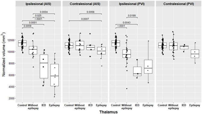
During a median follow-up time of 12.8 years [interquartile range (IQR): 10.8-17.3] in the AIS group and 12.5 years (IQR: 9.3-14.8) in the PVI group ( = 0.32), epilepsy developed in 10 children (34.5%) with AIS and in 4 (12.1%) children with PVI, = 0.036 [odds ratio (OR) = 3.8, 95%, confidence interval (CI): 1.04-14]. Epilepsy and interictal epileptiform discharges (IEDs) without clinical seizures were more often expressed in children with AIS ( = 16, 55%) than in children with PVI ( = 7, 21.2%), = 0.0057 (OR = 3.8 95% CI: 1.04-14). In the AIS group, the ipsilesional and contralesional thalamus, ipsilesional caudate nucleus, and nucleus accumbens were significantly smaller in children with epilepsy compared to children without epilepsy. In the PVI group, the ipsilesional thalamus, caudate nucleus, and nucleus accumbens were smaller in the pooled group of epilepsy plus IED alone compared to children without epilepsy.
In children with AIS, epilepsy or IED occurred more often compared to children with PVI. Both patients with AIS and PVI with severe damage to the basal ganglia and the thalamus have a higher risk of developing poststroke epilepsy and should be monitored more closely throughout childhood to initiate timely antiseizure medication and rehabilitation.
癫痫是围产期卒中最严重的后果之一。癫痫本身被认为是认知、语言和行为功能受损的一个风险因素。目前仍不清楚哪些儿童在围产期卒中后会发生癫痫。本研究旨在评估围产期卒中后儿童丘脑和基底神经节的体积与卒中后癫痫的关系。
这项随访研究纳入了29例围产期动脉缺血性卒中(AIS)患儿、33例疑似脑室周围静脉梗死(PVI)患儿以及46例年龄和性别匹配的健康对照。对4至18岁的儿童进行了磁共振成像检查,并采用分割法进行体积分析,以评估丘脑、尾状核、壳核、苍白球、海马、杏仁核和伏隔核的大小。
AIS组的中位随访时间为12.8年[四分位间距(IQR):10.8 - 17.3],PVI组为12.5年(IQR:9.3 - 14.8)(P = 0.32)。10例(34.5%)AIS患儿和4例(12.1%)PVI患儿发生了癫痫,P = 0.036[比值比(OR)= 3.8,95%置信区间(CI):1.04 - 14]。与PVI患儿(n = 7,21.2%)相比,癫痫和无临床发作的发作间期癫痫样放电(IEDs)在AIS患儿(n = 16,55%)中更常出现,P = 0.0057(OR = 3.8,95% CI:1.04 - 14)。在AIS组中,与无癫痫的患儿相比,癫痫患儿的患侧和对侧丘脑、患侧尾状核和伏隔核明显更小。在PVI组中,与无癫痫的患儿相比,仅癫痫加IEDs的合并组中患侧丘脑、尾状核和伏隔核更小。
与PVI患儿相比,AIS患儿癫痫或IEDs的发生更为频繁。基底神经节和丘脑严重受损的AIS和PVI患者发生卒中后癫痫的风险更高,在整个儿童期应进行更密切的监测,以便及时启动抗癫痫药物治疗和康复治疗。