Department of Immunology and Cell Biology, Faculty of Medicine and Health Sciences, Université de Sherbrooke, Sherbrooke, QC, Canada.
Department of Microbiology and Immunology, Keio University School of Medicine, Tokyo, Japan.
Front Immunol. 2023 Oct 4;14:1259246. doi: 10.3389/fimmu.2023.1259246. eCollection 2023.
Hepatic stellate cells (HSC) become activated, differentiate to myofibroblasts and produce extracellular fibrillar matrix during liver fibrosis. The hepatic fibrogenic response is orchestrated by reciprocal interactions between HSCs and macrophages and their secreted products. SOCS1 can regulate several cytokines and growth factors implicated in liver fibrosis. Here we investigated the role of SOCS1 in regulating HSC activation.
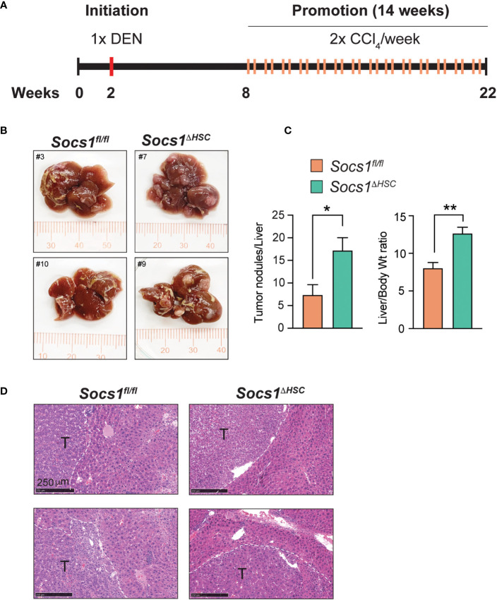
Mice lacking SOCS1 in HSCs () were generated by crossing and LratCre mice. Liver fibrosis was induced by carbon tetrachloride and evaluated by Sirius red staining, hydroxyproline content and immunostaining of myofibroblasts. Gene expression of pro-fibrogenic factors, cytokines, growth factors and chemokines were quantified by RT-qPCR. The phenotype and the numbers of intrahepatic leukocyte subsets were studied by flow cytometry. The impact of fibrosis on the development of diethyl nitrosamine-induced hepatocellular carcinoma was evaluated.
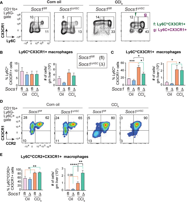
mice developed more severe liver fibrosis than control Socs1fl/fl mice that was characterized by increased collagen deposition and myofibroblast differentiation. mice showed a significant increase in the expression of smooth muscle actin, collagens, matrix metalloproteases, cytokines, growth factors and chemokines in the liver following fibrosis induction. The fibrotic livers of mice displayed heightened inflammatory cell infiltration with increased proportion and numbers of Ly6ChiCCR2+ pro-inflammatory macrophages. This macrophage population contained elevated numbers of CCR2+CX3CR1+ cells, suggesting impaired transition towards restorative macrophages. Fibrosis induction following exposure to diethyl nitrosamine resulted in more numerous and larger liver tumor nodules in mice than in mice.
Our findings indicate that (i) SOCS1 expression in HSCs is a critical to control liver fibrosis and development of hepatocaellular carcinoma, and (ii) attenuation of HSC activation by SOCS1 regulates pro-inflammatory macrophage recruitment and differentiation during liver fibrosis.
肝星状细胞(HSC)在肝纤维化过程中被激活、分化为肌成纤维细胞并产生细胞外纤维状基质。肝纤维化的纤维发生反应是由 HSC 和巨噬细胞及其分泌产物之间的相互作用协调的。SOCS1 可以调节几种细胞因子和生长因子,这些因子与肝纤维化有关。在这里,我们研究了 SOCS1 在调节 HSC 激活中的作用。
通过将 和 LratCre 小鼠杂交,生成缺乏 HSCs 中 SOCS1 的小鼠()。通过四氯化碳诱导肝纤维化,并通过天狼猩红染色、羟脯氨酸含量和肌成纤维细胞免疫染色评估。通过 RT-qPCR 定量分析促纤维化因子、细胞因子、生长因子和趋化因子的基因表达。通过流式细胞术研究肝内白细胞亚群的表型和数量。评估纤维化对二乙基亚硝胺诱导的肝细胞癌发展的影响。
与对照 Socs1fl/fl 小鼠相比, 小鼠发生更严重的肝纤维化,其特征为胶原沉积和肌成纤维细胞分化增加。纤维化诱导后, 小鼠肝脏中平滑肌肌动蛋白、胶原蛋白、基质金属蛋白酶、细胞因子、生长因子和趋化因子的表达显著增加。 小鼠纤维化肝脏的炎症细胞浸润明显增加,促炎巨噬细胞的比例和数量增加。这种巨噬细胞群体含有更多数量的 CCR2+CX3CR1+细胞,表明向修复性巨噬细胞的转变受损。暴露于二乙基亚硝胺后诱导纤维化导致 小鼠的肝肿瘤结节数量更多、更大。
我们的研究结果表明,(i)HSC 中的 SOCS1 表达对于控制肝纤维化和肝细胞癌的发生是至关重要的,(ii)SOCS1 对 HSC 激活的抑制调节了肝纤维化过程中促炎巨噬细胞的募集和分化。