• 文献检索
  • 文档翻译
  • 深度研究
  • 学术资讯
  • Suppr Zotero 插件Zotero 插件
  • 邀请有礼
  • 套餐&价格
  • 历史记录
应用&插件
Suppr Zotero 插件Zotero 插件浏览器插件Mac 客户端Windows 客户端微信小程序
定价
高级版会员购买积分包购买API积分包
服务
文献检索文档翻译深度研究API 文档MCP 服务
关于我们
关于 Suppr公司介绍联系我们用户协议隐私条款
关注我们

Suppr 超能文献

核心技术专利:CN118964589B侵权必究
粤ICP备2023148730 号-1Suppr @ 2026

文献检索

告别复杂PubMed语法,用中文像聊天一样搜索,搜遍4000万医学文献。AI智能推荐,让科研检索更轻松。

立即免费搜索

文件翻译

保留排版,准确专业,支持PDF/Word/PPT等文件格式,支持 12+语言互译。

免费翻译文档

深度研究

AI帮你快速写综述,25分钟生成高质量综述,智能提取关键信息,辅助科研写作。

立即免费体验

一种诱导型模型,用于遗传操作和肝内 PDGFRβ 表达纤维生成细胞的命运追踪。

An inducible model for genetic manipulation and fate-tracing of PDGFRβ-expressing fibrogenic cells in the liver.

机构信息

Department of Gastroenterology, Hepatology, Infectious Diseases and Endocrinology, Hannover Medical School, Carl-Neuberg-Str. 1, 30625, Hannover, Germany.

出版信息

Sci Rep. 2023 May 5;13(1):7322. doi: 10.1038/s41598-023-34353-y.

DOI:10.1038/s41598-023-34353-y
PMID:37147343
原文链接:https://pmc.ncbi.nlm.nih.gov/articles/PMC10162963/
Abstract

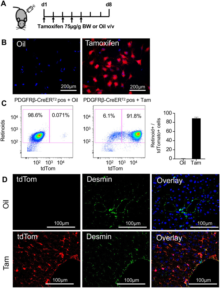
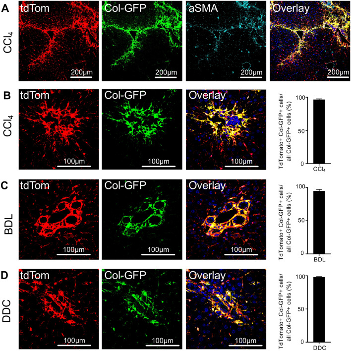
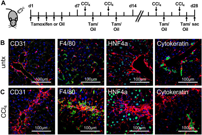
Myofibroblasts are the source of extracellular matrix protein during liver fibrogenesis. Fibroblasts, hepatic stellate cells (HSCs) and vascular smooth muscle cells are mesenchymal subpopulations in the liver that are characterized by the expression of PDGFRβ and contribute to the pool of these myofibroblasts. Conditional knockout models are important to better understand the function of specific liver cell populations including mesenchymal cells. While there is a limited number of constitutive mouse models for liver mesenchymal cell specific transgene expression, there is no established model for inducible gene targeting in HSCs or PDGFRβ-expressing mesenchymal cell populations in the liver. To address this, we investigated whether the tamoxifen inducible PDGFRβ-P2A-CreER mouse can be used as a reliable tool to specifically express transgens in liver mesenchymal cells. Our data demonstrate, that PDGFRβ-P2A-CreER specifically and efficiently marks over 90% of retinoid positive HSCs in healthy and fibrotic liver in mice upon tamoxifen injection, and that those cells give rise to Col1a1-expressing myofibroblasts in different models of liver fibrosis. Together with a negligible background recombination of only about 0.33%, this confirms that the PDGFRβ-P2A-CreER mouse is nearly as efficient as established constitutive LratCre and PDGFRβ-Cre mouse models for recombination in HSCs, and that it is a powerful model for mesenchymal liver cell studies that require an inducible Cre approach.

摘要

肌成纤维细胞是肝纤维化过程中细胞外基质蛋白的来源。成纤维细胞、肝星状细胞 (HSCs) 和血管平滑肌细胞是肝内的间充质亚群,其特征是表达 PDGFRβ,并有助于肌成纤维细胞池的形成。条件性敲除模型对于更好地理解特定的肝细胞群体(包括间充质细胞)的功能非常重要。虽然有数量有限的用于肝间充质细胞特异性转基因表达的组成型小鼠模型,但在 HSCs 或 PDGFRβ 表达的肝间充质细胞群体中没有建立用于诱导基因靶向的模型。为了解决这个问题,我们研究了是否可以将他莫昔芬诱导的 PDGFRβ-P2A-CreER 小鼠用作在肝间充质细胞中特异性表达转基因的可靠工具。我们的数据表明,在给予他莫昔芬注射后,PDGFRβ-P2A-CreER 特异性且有效地标记了健康和纤维化肝脏中超过 90%的视黄酸阳性 HSCs,并且这些细胞在不同的肝纤维化模型中产生表达 Col1a1 的肌成纤维细胞。与仅约 0.33%的可忽略不计的背景重组相结合,这证实了 PDGFRβ-P2A-CreER 小鼠在 HSCs 中的重组效率几乎与已建立的组成型 LratCre 和 PDGFRβ-Cre 小鼠模型一样高效,并且它是一种用于需要诱导 Cre 方法的间充质肝细胞研究的强大模型。

https://cdn.ncbi.nlm.nih.gov/pmc/blobs/f042/10162963/ebd32ce6b011/41598_2023_34353_Fig3_HTML.jpg
https://cdn.ncbi.nlm.nih.gov/pmc/blobs/f042/10162963/fac3e01bc611/41598_2023_34353_Fig1_HTML.jpg
https://cdn.ncbi.nlm.nih.gov/pmc/blobs/f042/10162963/26e77f3b9454/41598_2023_34353_Fig2_HTML.jpg
https://cdn.ncbi.nlm.nih.gov/pmc/blobs/f042/10162963/ebd32ce6b011/41598_2023_34353_Fig3_HTML.jpg
https://cdn.ncbi.nlm.nih.gov/pmc/blobs/f042/10162963/fac3e01bc611/41598_2023_34353_Fig1_HTML.jpg
https://cdn.ncbi.nlm.nih.gov/pmc/blobs/f042/10162963/26e77f3b9454/41598_2023_34353_Fig2_HTML.jpg
https://cdn.ncbi.nlm.nih.gov/pmc/blobs/f042/10162963/ebd32ce6b011/41598_2023_34353_Fig3_HTML.jpg

相似文献

1
An inducible model for genetic manipulation and fate-tracing of PDGFRβ-expressing fibrogenic cells in the liver.一种诱导型模型,用于遗传操作和肝内 PDGFRβ 表达纤维生成细胞的命运追踪。
Sci Rep. 2023 May 5;13(1):7322. doi: 10.1038/s41598-023-34353-y.
2
Hepatic stellate cells in zone 1 engage in capillarization rather than myofibroblast formation in murine liver fibrosis.1 区的肝星状细胞在肝纤维化中发生毛细血管化,而不是肌成纤维细胞形成。
Sci Rep. 2024 Aug 13;14(1):18840. doi: 10.1038/s41598-024-69898-z.
3
PDGFRβ-P2A-CreER mice: a genetic tool to target pericytes in angiogenesis.PDGFRβ-P2A-CreER 小鼠:一种用于血管生成中靶向周细胞的遗传工具。
Angiogenesis. 2017 Nov;20(4):655-662. doi: 10.1007/s10456-017-9570-9. Epub 2017 Jul 27.
4
Genetic labeling does not detect epithelial-to-mesenchymal transition of cholangiocytes in liver fibrosis in mice.遗传标记不能检测到小鼠肝纤维化中胆管细胞的上皮间质转化。
Gastroenterology. 2010 Sep;139(3):987-98. doi: 10.1053/j.gastro.2010.05.005. Epub 2010 Jun 20.
5
Perivenous Stellate Cells Are the Main Source of Myofibroblasts and Cancer-Associated Fibroblasts Formed After Chronic Liver Injuries.慢性肝损伤后形成的肌成纤维细胞和癌相关成纤维细胞主要来源于肝窦周星形细胞。
Hepatology. 2021 Sep;74(3):1578-1594. doi: 10.1002/hep.31848.
6
Mesodermal mesenchymal cells give rise to myofibroblasts, but not epithelial cells, in mouse liver injury.中胚层间充质细胞在小鼠肝损伤中产生肌成纤维细胞,但不产生上皮细胞。
Hepatology. 2014 Jul;60(1):311-22. doi: 10.1002/hep.27035. Epub 2014 Apr 28.
7
Single Cell RNA Sequencing Identifies Subsets of Hepatic Stellate Cells and Myofibroblasts in Liver Fibrosis.单细胞 RNA 测序鉴定肝纤维化中肝星状细胞和肌成纤维细胞亚群。
Cells. 2019 May 24;8(5):503. doi: 10.3390/cells8050503.
8
Hedgehog-YAP Signaling Pathway Regulates Glutaminolysis to Control Activation of Hepatic Stellate Cells.刺猬-YAP 信号通路调控谷氨酰胺代谢以控制肝星状细胞的激活。
Gastroenterology. 2018 Apr;154(5):1465-1479.e13. doi: 10.1053/j.gastro.2017.12.022. Epub 2018 Jan 3.
9
Mesothelial cells give rise to hepatic stellate cells and myofibroblasts via mesothelial-mesenchymal transition in liver injury.在肝损伤中,间皮细胞通过间皮-间质转化产生肝星状细胞和肌成纤维细胞。
Proc Natl Acad Sci U S A. 2013 Feb 5;110(6):2324-9. doi: 10.1073/pnas.1214136110. Epub 2013 Jan 23.
10
P300 Acetyltransferase Mediates Stiffness-Induced Activation of Hepatic Stellate Cells Into Tumor-Promoting Myofibroblasts.P300 乙酰转移酶介导肝星状细胞硬度诱导激活为促肿瘤肌成纤维细胞。
Gastroenterology. 2018 Jun;154(8):2209-2221.e14. doi: 10.1053/j.gastro.2018.02.015. Epub 2018 Feb 15.

引用本文的文献

1
Molecular mechanisms behind the inhibitory effects of ginsenoside Rg3 on hepatic fibrosis: a review.人参皂苷Rg3对肝纤维化抑制作用的分子机制综述
Arch Toxicol. 2025 Feb;99(2):541-561. doi: 10.1007/s00204-024-03941-w. Epub 2024 Dec 27.
2
Regulation of High-fat Diet-induced Liver Fibrosis by SOCS1 Expression in Hepatic Stellate Cells.肝星状细胞中SOCS1表达对高脂饮食诱导的肝纤维化的调控
J Clin Exp Hepatol. 2024 Jan-Feb;14(1):101280. doi: 10.1016/j.jceh.2023.09.001. Epub 2023 Sep 9.
3
Hepatic stellate cell-intrinsic role of SOCS1 in controlling hepatic fibrogenic response and the pro-inflammatory macrophage compartment during liver fibrosis.

本文引用的文献

1
Opposing roles of hepatic stellate cell subpopulations in hepatocarcinogenesis.肝星状细胞亚群在肝癌发生中的相反作用。
Nature. 2022 Oct;610(7931):356-365. doi: 10.1038/s41586-022-05289-6. Epub 2022 Oct 5.
2
Portal fibroblasts with mesenchymal stem cell features form a reservoir of proliferative myofibroblasts in liver fibrosis.具有间充质干细胞特征的肝门成纤维细胞在肝纤维化过程中形成增殖性肌成纤维细胞的储备库。
Hepatology. 2022 Nov;76(5):1360-1375. doi: 10.1002/hep.32456. Epub 2022 Mar 29.
3
Emerging synthetic drugs for the treatment of liver cirrhosis.
SOCS1 在控制肝纤维化过程中肝星状细胞固有作用及肝纤维化时促炎性巨噬细胞区室。
Front Immunol. 2023 Oct 4;14:1259246. doi: 10.3389/fimmu.2023.1259246. eCollection 2023.
新兴的治疗肝硬化的合成药物。
Expert Opin Emerg Drugs. 2021 Jun;26(2):149-163. doi: 10.1080/14728214.2021.1918099. Epub 2021 Apr 26.
4
-regulated activation of hepatic stellate cells and liver fibrosis by TGF-β signaling.TGF-β 信号调控肝星状细胞激活和肝纤维化。
Am J Physiol Gastrointest Liver Physiol. 2021 May 1;320(5):G720-G728. doi: 10.1152/ajpgi.00310.2020. Epub 2021 Mar 17.
5
Fibrosis: from mechanisms to medicines.纤维化:从机制到药物
Nature. 2020 Nov;587(7835):555-566. doi: 10.1038/s41586-020-2938-9. Epub 2020 Nov 25.
6
Hepatic stellate cell hypertrophy is associated with metabolic liver fibrosis.肝星状细胞肥大与代谢性肝纤维化有关。
Sci Rep. 2020 Mar 2;10(1):3850. doi: 10.1038/s41598-020-60615-0.
7
Single-Cell Transcriptomics Uncovers Zonation of Function in the Mesenchyme during Liver Fibrosis.单细胞转录组学揭示肝纤维化过程中间质功能的分区。
Cell Rep. 2019 Nov 12;29(7):1832-1847.e8. doi: 10.1016/j.celrep.2019.10.024.
8
Global, regional, and national age-sex-specific mortality for 282 causes of death in 195 countries and territories, 1980-2017: a systematic analysis for the Global Burden of Disease Study 2017.全球、区域和国家按年龄、性别和死因分类的死亡率,195 个国家和地区,1980-2017 年:2017 年全球疾病负担研究的系统分析。
Lancet. 2018 Nov 10;392(10159):1736-1788. doi: 10.1016/S0140-6736(18)32203-7. Epub 2018 Nov 8.
9
Stellate Cells in Tissue Repair, Inflammation, and Cancer.星状细胞在组织修复、炎症和癌症中的作用。
Annu Rev Cell Dev Biol. 2018 Oct 6;34:333-355. doi: 10.1146/annurev-cellbio-100617-062855. Epub 2018 Jul 20.
10
Desmin- and vimentin-mediated hepatic stellate cell-targeting radiotracer Tc-GlcNAc-PEI for liver fibrosis imaging with SPECT.使用 SPECT 对肝纤维化进行成像的基于 Desmin 和 Vimentin 的肝星状细胞靶向放射性示踪剂 Tc-GlcNAc-PEI。
Theranostics. 2018 Feb 2;8(5):1340-1349. doi: 10.7150/thno.22806. eCollection 2018.