• 文献检索
  • 文档翻译
  • 深度研究
  • 学术资讯
  • Suppr Zotero 插件Zotero 插件
  • 邀请有礼
  • 套餐&价格
  • 历史记录
应用&插件
Suppr Zotero 插件Zotero 插件浏览器插件Mac 客户端Windows 客户端微信小程序
定价
高级版会员购买积分包购买API积分包
服务
文献检索文档翻译深度研究API 文档MCP 服务
关于我们
关于 Suppr公司介绍联系我们用户协议隐私条款
关注我们

Suppr 超能文献

核心技术专利:CN118964589B侵权必究
粤ICP备2023148730 号-1Suppr @ 2026

文献检索

告别复杂PubMed语法,用中文像聊天一样搜索,搜遍4000万医学文献。AI智能推荐,让科研检索更轻松。

立即免费搜索

文件翻译

保留排版,准确专业,支持PDF/Word/PPT等文件格式,支持 12+语言互译。

免费翻译文档

深度研究

AI帮你快速写综述,25分钟生成高质量综述,智能提取关键信息,辅助科研写作。

立即免费体验

单细胞分析联合谱系分析揭示迷走神经和骶神经嵴对发育中肠神经的贡献。

Single-cell profiling coupled with lineage analysis reveals vagal and sacral neural crest contributions to the developing enteric nervous system.

机构信息

Division of Biology and Biological Engineering, California Institute of Technology, Pasadena, United States.

出版信息

Elife. 2023 Oct 25;12:e79156. doi: 10.7554/eLife.79156.

DOI:10.7554/eLife.79156
PMID:37877560
原文链接:https://pmc.ncbi.nlm.nih.gov/articles/PMC10627514/
Abstract

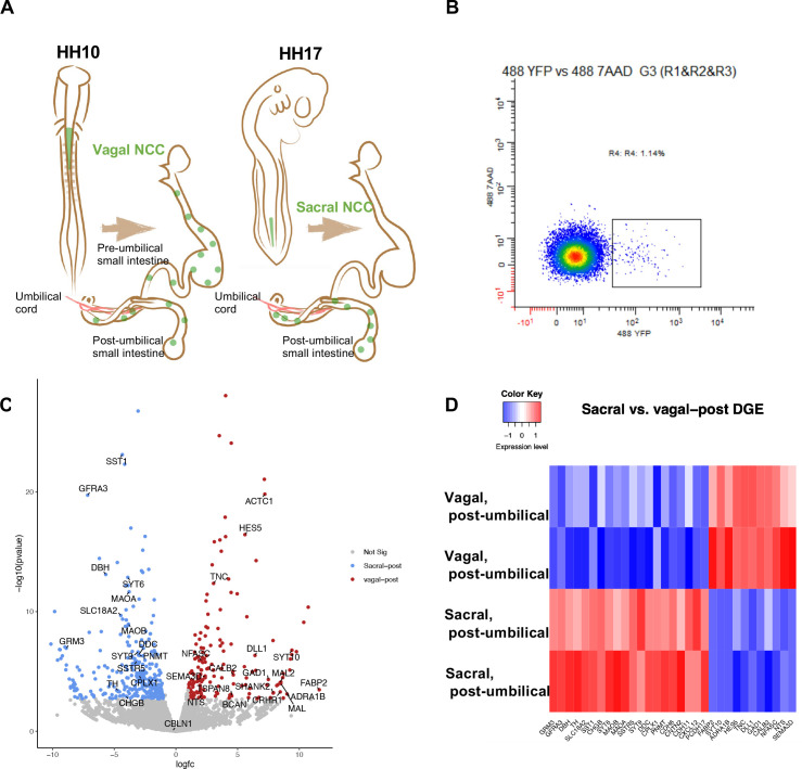
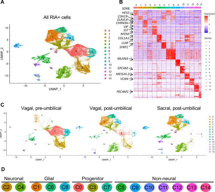
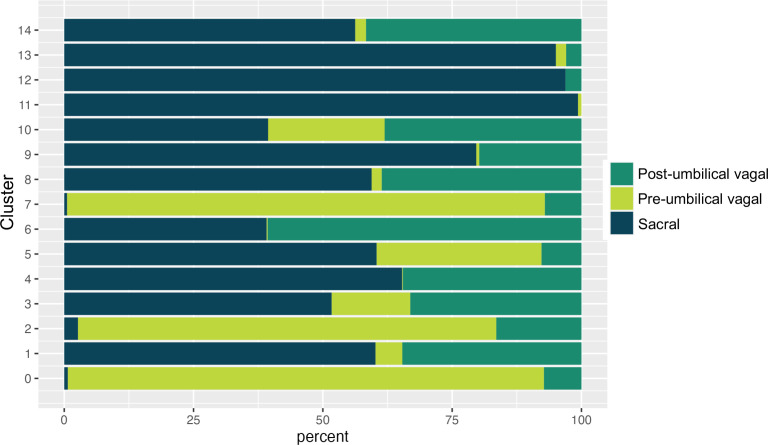
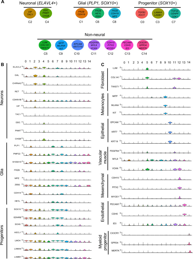
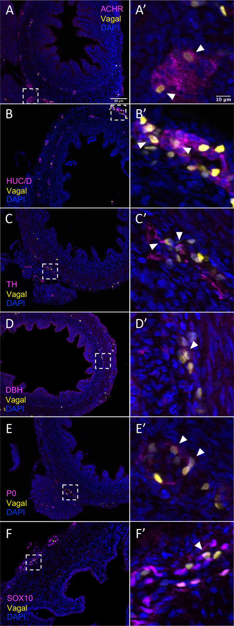
During development, much of the enteric nervous system (ENS) arises from the vagal neural crest that emerges from the caudal hindbrain and colonizes the entire gastrointestinal tract. However, a second ENS contribution comes from the sacral neural crest that arises in the caudal neural tube and populates the post-umbilical gut. By coupling single-cell transcriptomics with axial-level-specific lineage tracing in avian embryos, we compared the contributions of embryonic vagal and sacral neural crest cells to the chick ENS and the associated peripheral ganglia (Nerve of Remak and pelvic plexuses). At embryonic day (E) 10, the two neural crest populations form overlapping subsets of neuronal and glia cell types. Surprisingly, the post-umbilical vagal neural crest much more closely resembles the sacral neural crest than the pre-umbilical vagal neural crest. However, some differences in cluster types were noted between vagal and sacral derived cells. Notably, RNA trajectory analysis suggests that the vagal neural crest maintains a neuronal/glial progenitor pool, whereas this cluster is depleted in the E10 sacral neural crest which instead has numerous enteric glia. The present findings reveal sacral neural crest contributions to the hindgut and associated peripheral ganglia and highlight the potential influence of the local environment and/or developmental timing in differentiation of neural crest-derived cells in the developing ENS.

摘要

在发育过程中,大部分肠神经系统 (ENS) 来自于迷走神经嵴,它起源于后脑尾部并殖民整个胃肠道。然而,ENS 的第二个来源是来自于尾侧神经管的骶神经嵴,它在尾侧神经管中产生并填充了脐带后的肠道。通过将单细胞转录组学与禽类胚胎的轴向水平特异性谱系追踪相结合,我们比较了胚胎迷走神经和骶神经嵴细胞对鸡 ENS 及其相关的周围神经节(Remak 神经和骨盆丛)的贡献。在胚胎第 10 天 (E10),两个神经嵴群体形成了重叠的神经元和神经胶质细胞类型子集。令人惊讶的是,脐带后的迷走神经嵴比脐带前的迷走神经嵴更类似于骶神经嵴。然而,在迷走神经和骶神经来源的细胞之间观察到一些簇类型的差异。值得注意的是,RNA 轨迹分析表明,迷走神经嵴保持着神经元/神经胶质祖细胞池,而这个簇在 E10 骶神经嵴中被耗尽,而后者有许多肠神经胶质细胞。本研究结果揭示了骶神经嵴对后肠和相关周围神经节的贡献,并强调了局部环境和/或发育时间在分化发育中的 ENS 中的神经嵴衍生细胞中的潜在影响。

https://cdn.ncbi.nlm.nih.gov/pmc/blobs/f652/10627514/dca484d3c868/elife-79156-fig8-figsupp1.jpg
https://cdn.ncbi.nlm.nih.gov/pmc/blobs/f652/10627514/f005553c398c/elife-79156-fig1.jpg
https://cdn.ncbi.nlm.nih.gov/pmc/blobs/f652/10627514/e2c2cf7f7823/elife-79156-fig1-figsupp1.jpg
https://cdn.ncbi.nlm.nih.gov/pmc/blobs/f652/10627514/b2c4df5dc51f/elife-79156-fig1-figsupp2.jpg
https://cdn.ncbi.nlm.nih.gov/pmc/blobs/f652/10627514/cd1bbd7e0fe3/elife-79156-fig2.jpg
https://cdn.ncbi.nlm.nih.gov/pmc/blobs/f652/10627514/9fad2252b4a0/elife-79156-fig2-figsupp1.jpg
https://cdn.ncbi.nlm.nih.gov/pmc/blobs/f652/10627514/b79ac566027d/elife-79156-fig3.jpg
https://cdn.ncbi.nlm.nih.gov/pmc/blobs/f652/10627514/403a07d70035/elife-79156-fig4.jpg
https://cdn.ncbi.nlm.nih.gov/pmc/blobs/f652/10627514/193dd313f365/elife-79156-fig4-figsupp1.jpg
https://cdn.ncbi.nlm.nih.gov/pmc/blobs/f652/10627514/5bded85a4873/elife-79156-fig4-figsupp2.jpg
https://cdn.ncbi.nlm.nih.gov/pmc/blobs/f652/10627514/22c526400ecc/elife-79156-fig5.jpg
https://cdn.ncbi.nlm.nih.gov/pmc/blobs/f652/10627514/a94c1bd91af2/elife-79156-fig5-figsupp1.jpg
https://cdn.ncbi.nlm.nih.gov/pmc/blobs/f652/10627514/445881939a75/elife-79156-fig5-figsupp2.jpg
https://cdn.ncbi.nlm.nih.gov/pmc/blobs/f652/10627514/8c7dafbcb30d/elife-79156-fig6.jpg
https://cdn.ncbi.nlm.nih.gov/pmc/blobs/f652/10627514/4de9fee6c577/elife-79156-fig6-figsupp1.jpg
https://cdn.ncbi.nlm.nih.gov/pmc/blobs/f652/10627514/595759cac1cb/elife-79156-fig7.jpg
https://cdn.ncbi.nlm.nih.gov/pmc/blobs/f652/10627514/1b3f31f1e485/elife-79156-fig8.jpg
https://cdn.ncbi.nlm.nih.gov/pmc/blobs/f652/10627514/dca484d3c868/elife-79156-fig8-figsupp1.jpg

相似文献

1
Single-cell profiling coupled with lineage analysis reveals vagal and sacral neural crest contributions to the developing enteric nervous system.单细胞分析联合谱系分析揭示迷走神经和骶神经嵴对发育中肠神经的贡献。
Elife. 2023 Oct 25;12:e79156. doi: 10.7554/eLife.79156.
2
Sacral neural crest cells colonise aganglionic hindgut in vivo but fail to compensate for lack of enteric ganglia.骶神经嵴细胞在体内定殖于无神经节的后肠,但无法弥补肠神经节的缺失。
Dev Biol. 2000 Mar 1;219(1):30-43. doi: 10.1006/dbio.1999.9592.
3
In ovo transplantation of enteric nervous system precursors from vagal to sacral neural crest results in extensive hindgut colonisation.将迷走神经至骶神经嵴的肠神经系统前体进行卵内移植,会导致后肠广泛定植。
Development. 2002 Jun;129(12):2785-96. doi: 10.1242/dev.129.12.2785.
4
The sacral neural crest contributes neurons and glia to the post-umbilical gut: spatiotemporal analysis of the development of the enteric nervous system.骶神经嵴为脐后肠道提供神经元和神经胶质细胞:肠神经系统发育的时空分析。
Development. 1998 Nov;125(21):4335-47. doi: 10.1242/dev.125.21.4335.
5
Enteric nervous system development: analysis of the selective developmental potentialities of vagal and sacral neural crest cells using quail-chick chimeras.肠神经系统发育:利用鹌鹑-鸡嵌合体分析迷走神经和骶神经嵴细胞的选择性发育潜能
Anat Rec. 2001 Jan 1;262(1):16-28. doi: 10.1002/1097-0185(20010101)262:1<16::AID-AR1007>3.0.CO;2-O.
6
Enteric nervous system specific deletion of Foxd3 disrupts glial cell differentiation and activates compensatory enteric progenitors.肠神经系统特异性敲除 Foxd3 会破坏神经胶质细胞分化并激活代偿性肠前体细胞。
Dev Biol. 2012 Mar 15;363(2):373-87. doi: 10.1016/j.ydbio.2012.01.003. Epub 2012 Jan 12.
7
Analysis of the sacral neural crest cell contribution to the hindgut enteric nervous system in the mouse embryo.分析鼠胚骶神经嵴细胞对后肠肠神经嵴细胞的贡献。
Gastroenterology. 2011 Sep;141(3):992-1002.e1-6. doi: 10.1053/j.gastro.2011.06.002. Epub 2011 Jun 13.
8
Lumbo-sacral neural crest contributes to the avian enteric nervous system independently of vagal neural crest.腰骶神经嵴独立于迷走神经嵴对鸟类肠神经系统有贡献。
Dev Dyn. 2000 Jul;218(3):525-30. doi: 10.1002/1097-0177(200007)218:3<525::AID-DVDY1003>3.0.CO;2-7.
9
Colonization of the bowel by neural crest-derived cells re-migrating from foregut backtransplanted to vagal or sacral regions of host embryos.从宿主胚胎的迷走神经或骶骨区域回植到前肠的神经嵴衍生细胞重新迁移至肠道并定殖。
Dev Dyn. 1993 Mar;196(3):217-33. doi: 10.1002/aja.1001960308.
10
Critical numbers of neural crest cells are required in the pathways from the neural tube to the foregut to ensure complete enteric nervous system formation.从神经管到前肠的通路中需要关键数量的神经嵴细胞,以确保完整的肠神经系统形成。
Development. 2008 May;135(9):1681-91. doi: 10.1242/dev.017418. Epub 2008 Apr 2.

引用本文的文献

1
Whole-Gut Spatial Genomic Analysis Reveals Molecular Regionalization of the Differentiating Zebrafish Enteric Nervous System.全肠道空间基因组分析揭示斑马鱼分化中肠神经系统的分子区域化。
FASEB J. 2025 Sep 15;39(17):e71019. doi: 10.1096/fj.202501470R.
2
genes affect neuronal differentiation in the developing zebrafish enteric nervous system.基因影响发育中的斑马鱼肠道神经系统中的神经元分化。
Proc Natl Acad Sci U S A. 2025 Aug 19;122(33):e2510548122. doi: 10.1073/pnas.2510548122. Epub 2025 Aug 11.
3
Wnt signaling pathway and retinoic acid signaling pathway involved in delamination and migration of chicken trunk NCCs and contributing to HVP phenotype.

本文引用的文献

1
A branching model of lineage differentiation underpinning the neurogenic potential of enteric glia.分支模型的谱系分化为基础的神经发生潜力的肠胶质细胞。
Nat Commun. 2023 Sep 22;14(1):5904. doi: 10.1038/s41467-023-41492-3.
2
hPSC-derived sacral neural crest enables rescue in a severe model of Hirschsprung's disease.人多能干细胞源性荐骨神经嵴细胞可挽救严重先天性巨结肠疾病模型。
Cell Stem Cell. 2023 Mar 2;30(3):264-282.e9. doi: 10.1016/j.stem.2023.02.003.
3
CellRank for directed single-cell fate mapping.细胞排序用于有向单细胞命运图谱绘制。
Wnt信号通路和视黄酸信号通路参与鸡胚躯干神经嵴细胞的分层和迁移,并促成泄殖腔后肠表型。
Poult Sci. 2025 Apr 3;104(6):105114. doi: 10.1016/j.psj.2025.105114.
4
Ancient emergence of neuronal heterogeneity in the enteric nervous system of jawless vertebrates.无颌脊椎动物肠神经系统中神经元异质性的古老起源。
Dev Biol. 2025 Apr;520:117-124. doi: 10.1016/j.ydbio.2024.12.020. Epub 2025 Jan 3.
5
Spatiotemporal dynamics of the developing zebrafish enteric nervous system at the whole-organ level.斑马鱼发育中的肠道神经系统在全器官水平的时空动态变化。
Dev Cell. 2025 Feb 24;60(4):613-629.e6. doi: 10.1016/j.devcel.2024.11.006. Epub 2024 Dec 5.
6
Human enteric nervous system progenitor transplantation improves functional responses in Hirschsprung disease patient-derived tissue.人肠道神经系统祖细胞移植改善了先天性巨结肠病患者来源组织的功能反应。
Gut. 2024 Aug 8;73(9):1441-1453. doi: 10.1136/gutjnl-2023-331532.
7
CXCR4 and CXCL12 signaling regulates the development of extrinsic innervation to the colorectum.CXCR4 和 CXCL12 信号调节直肠外神经支配的发育。
Development. 2023 Apr 15;150(8). doi: 10.1242/dev.201289. Epub 2023 Apr 20.
Nat Methods. 2022 Feb;19(2):159-170. doi: 10.1038/s41592-021-01346-6. Epub 2022 Jan 13.
4
Roles of Enteric Neural Stem Cell Niche and Enteric Nervous System Development in Hirschsprung Disease.肠神经干细胞龛和肠神经系统发育在先天性巨结肠病中的作用。
Int J Mol Sci. 2021 Sep 7;22(18):9659. doi: 10.3390/ijms22189659.
5
Single-cell transcriptomic landscape of cardiac neural crest cell derivatives during development.心脏神经嵴细胞衍生物在发育过程中的单细胞转录组图谱。
EMBO Rep. 2021 Nov 4;22(11):e52389. doi: 10.15252/embr.202152389. Epub 2021 Sep 27.
6
Direct Interaction of Sox10 With Cadherin-19 Mediates Early Sacral Neural Crest Cell Migration: Implications for Enteric Nervous System Development Defects.Sox10 与钙黏蛋白-19 的直接相互作用介导早期荐颅神经嵴细胞迁移:对肠神经系统发育缺陷的影响。
Gastroenterology. 2022 Jan;162(1):179-192.e11. doi: 10.1053/j.gastro.2021.08.029. Epub 2021 Aug 21.
7
5-HT3 Signaling Alters Development of Sacral Neural Crest Derivatives That Innervate the Lower Urinary Tract.5-HT3 信号转导改变支配下尿路的骶神经嵴衍生物的发育。
Int J Mol Sci. 2021 Jun 25;22(13):6838. doi: 10.3390/ijms22136838.
8
Altered sacral neural crest development in Pax3 spina bifida mutants underlies deficits of bladder innervation and function.Pax3 脊柱裂突变体中骶神经嵴发育改变是膀胱神经支配和功能缺陷的基础。
Dev Biol. 2021 Aug;476:173-188. doi: 10.1016/j.ydbio.2021.03.024. Epub 2021 Apr 9.
9
Clonal analysis and dynamic imaging identify multipotency of individual Gallus gallus caudal hindbrain neural crest cells toward cardiac and enteric fates.克隆分析和动态成像鉴定了鸡尾部后脑神经嵴细胞向心脏和肠命运的多能性。
Nat Commun. 2021 Mar 25;12(1):1894. doi: 10.1038/s41467-021-22146-8.
10
An atlas of neural crest lineages along the posterior developing zebrafish at single-cell resolution.单细胞分辨率下沿斑马鱼后发育轴的神经嵴谱系图谱。
Elife. 2021 Feb 16;10:e60005. doi: 10.7554/eLife.60005.