• 文献检索
  • 文档翻译
  • 深度研究
  • 学术资讯
  • Suppr Zotero 插件Zotero 插件
  • 邀请有礼
  • 套餐&价格
  • 历史记录
应用&插件
Suppr Zotero 插件Zotero 插件浏览器插件Mac 客户端Windows 客户端微信小程序
定价
高级版会员购买积分包购买API积分包
服务
文献检索文档翻译深度研究API 文档MCP 服务
关于我们
关于 Suppr公司介绍联系我们用户协议隐私条款
关注我们

Suppr 超能文献

核心技术专利:CN118964589B侵权必究
粤ICP备2023148730 号-1Suppr @ 2026

文献检索

告别复杂PubMed语法,用中文像聊天一样搜索,搜遍4000万医学文献。AI智能推荐,让科研检索更轻松。

立即免费搜索

文件翻译

保留排版,准确专业,支持PDF/Word/PPT等文件格式,支持 12+语言互译。

免费翻译文档

深度研究

AI帮你快速写综述,25分钟生成高质量综述,智能提取关键信息,辅助科研写作。

立即免费体验

使用快照高光谱成像(sHSI)相机和机器学习分类器进行小儿脑组织分割

Pediatric Brain Tissue Segmentation Using a Snapshot Hyperspectral Imaging (sHSI) Camera and Machine Learning Classifier.

作者信息

Kifle Naomi, Teti Saige, Ning Bo, Donoho Daniel A, Katz Itai, Keating Robert, Cha Richard Jaepyeong

机构信息

Sheikh Zayed Institute for Pediatric Surgical Innovation, Children's National Hospital, Washington, DC 20010, USA.

Department of Neurosurgery, Children's National Hospital, Washington, DC 20010, USA.

出版信息

Bioengineering (Basel). 2023 Oct 13;10(10):1190. doi: 10.3390/bioengineering10101190.

DOI:10.3390/bioengineering10101190
PMID:37892919
原文链接:https://pmc.ncbi.nlm.nih.gov/articles/PMC10603997/
Abstract

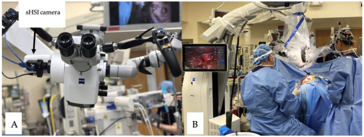
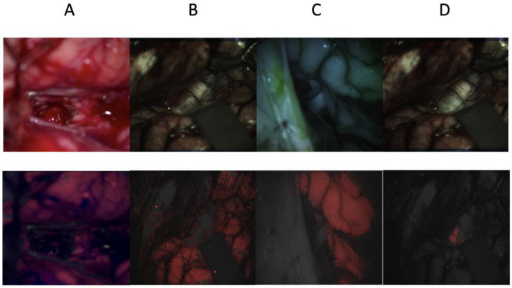
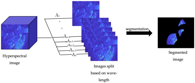
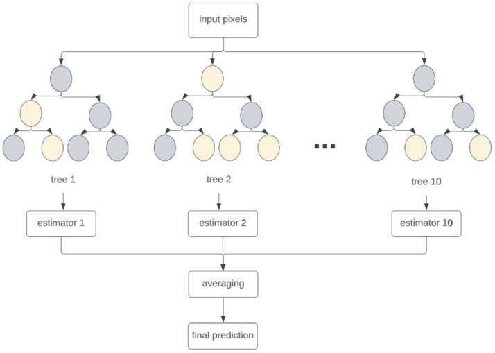
Pediatric brain tumors are the second most common type of cancer, accounting for one in four childhood cancer types. Brain tumor resection surgery remains the most common treatment option for brain cancer. While assessing tumor margins intraoperatively, surgeons must send tissue samples for biopsy, which can be time-consuming and not always accurate or helpful. Snapshot hyperspectral imaging (sHSI) cameras can capture scenes beyond the human visual spectrum and provide real-time guidance where we aim to segment healthy brain tissues from lesions on pediatric patients undergoing brain tumor resection. With the institutional research board approval, Pro00011028, 139 red-green-blue (RGB), 279 visible, and 85 infrared sHSI data were collected from four subjects with the system integrated into an operating microscope. A random forest classifier was used for data analysis. The RGB, infrared sHSI, and visible sHSI models achieved average intersection of unions (IoUs) of 0.76, 0.59, and 0.57, respectively, while the tumor segmentation achieved a specificity of 0.996, followed by the infrared HSI and visible HSI models at 0.93 and 0.91, respectively. Despite the small dataset considering pediatric cases, our research leveraged sHSI technology and successfully segmented healthy brain tissues from lesions with a high specificity during pediatric brain tumor resection procedures.

摘要

小儿脑肿瘤是第二常见的癌症类型,占儿童癌症类型的四分之一。脑肿瘤切除手术仍然是脑癌最常见的治疗选择。在术中评估肿瘤边界时,外科医生必须送检组织样本进行活检,这可能很耗时,而且并不总是准确或有帮助。快照高光谱成像(sHSI)相机可以捕捉超出人类视觉光谱范围的场景,并在我们旨在从接受脑肿瘤切除的儿科患者的病变中分割健康脑组织时提供实时指导。经机构研究委员会批准(Pro00011028),使用集成到手术显微镜中的系统从四名受试者收集了139个红绿蓝(RGB)、279个可见光和85个红外sHSI数据。使用随机森林分类器进行数据分析。RGB、红外sHSI和可见光sHSI模型的平均交并比(IoU)分别为0.76、0.59和0.57,而肿瘤分割的特异性为0.996,其次是红外HSI和可见光HSI模型,分别为0.93和0.91。尽管考虑到儿科病例的数据集较小,但我们的研究利用了sHSI技术,并在小儿脑肿瘤切除手术过程中成功地以高特异性从病变中分割出健康脑组织。

https://cdn.ncbi.nlm.nih.gov/pmc/blobs/e0bc/10603997/dbe9a85aee5e/bioengineering-10-01190-g005.jpg
https://cdn.ncbi.nlm.nih.gov/pmc/blobs/e0bc/10603997/78d089def220/bioengineering-10-01190-g001.jpg
https://cdn.ncbi.nlm.nih.gov/pmc/blobs/e0bc/10603997/c5593ed6bc71/bioengineering-10-01190-g002.jpg
https://cdn.ncbi.nlm.nih.gov/pmc/blobs/e0bc/10603997/01f0233ed376/bioengineering-10-01190-g003.jpg
https://cdn.ncbi.nlm.nih.gov/pmc/blobs/e0bc/10603997/dd614fdbc737/bioengineering-10-01190-g004.jpg
https://cdn.ncbi.nlm.nih.gov/pmc/blobs/e0bc/10603997/dbe9a85aee5e/bioengineering-10-01190-g005.jpg

相似文献

1
Pediatric Brain Tissue Segmentation Using a Snapshot Hyperspectral Imaging (sHSI) Camera and Machine Learning Classifier.使用快照高光谱成像(sHSI)相机和机器学习分类器进行小儿脑组织分割
Bioengineering (Basel). 2023 Oct 13;10(10):1190. doi: 10.3390/bioengineering10101190.
2
Spectral analysis comparison of pushbroom and snapshot hyperspectral cameras for brain tissues and chromophore identification.推帚式和快照式高光谱相机在脑组织和发色团识别中的光谱分析比较。
J Biomed Opt. 2024 Sep;29(9):093510. doi: 10.1117/1.JBO.29.9.093510. Epub 2024 Sep 24.
3
Toward assessment of resection margins using hyperspectral diffuse reflection imaging (400-1,700 nm) during tongue cancer surgery.利用舌癌手术期间的高光谱漫反射成像(400-1700nm)评估切缘。
Lasers Surg Med. 2020 Jul;52(6):496-502. doi: 10.1002/lsm.23161. Epub 2019 Sep 15.
4
Nerve Detection and Visualization Using Hyperspectral Imaging for Surgical Guidance.利用高光谱成像进行神经检测与可视化以辅助手术导航
Proc SPIE Int Soc Opt Eng. 2024 Feb;12930. doi: 10.1117/12.3008470. Epub 2024 Apr 2.
5
Estimation of tissue oxygen saturation from RGB images and sparse hyperspectral signals based on conditional generative adversarial network.基于条件生成对抗网络的 RGB 图像和稀疏高光谱信号的组织氧饱和度估计。
Int J Comput Assist Radiol Surg. 2019 Jun;14(6):987-995. doi: 10.1007/s11548-019-01940-2. Epub 2019 Mar 21.
6
Hyperspectral imaging for tumor segmentation on pigmented skin lesions.高光谱成像在色素皮损肿瘤分割中的应用。
J Biomed Opt. 2022 Oct;27(10). doi: 10.1117/1.JBO.27.10.106007.
7
Augmented reality visualization of hyperspectral imaging classifications for image-guided brain tumor phantom resection.用于图像引导脑肿瘤模型切除的高光谱成像分类的增强现实可视化。
Proc SPIE Int Soc Opt Eng. 2020 Feb;11315. doi: 10.1117/12.2549041. Epub 2020 Mar 16.
8
Deep learning approach for hyperspectral image demosaicking, spectral correction and high-resolution RGB reconstruction.用于高光谱图像去马赛克、光谱校正和高分辨率RGB重建的深度学习方法。
Comput Methods Biomech Biomed Eng Imaging Vis. 2021 Nov 30;10(4):409-417. doi: 10.1080/21681163.2021.1997646. eCollection 2022.
9
Automated Tumor Segmentation and Brain Tissue Extraction from Multiparametric MRI of Pediatric Brain Tumors: A Multi-Institutional Study.从儿科脑肿瘤多参数磁共振成像中自动进行肿瘤分割和脑组织提取:一项多机构研究。
medRxiv. 2023 Jan 11:2023.01.02.22284037. doi: 10.1101/2023.01.02.22284037.
10
Validation of two techniques for intraoperative hyperspectral human tissue determination.两种术中高光谱人体组织测定技术的验证
J Med Imaging (Bellingham). 2020 Nov;7(6):065001. doi: 10.1117/1.JMI.7.6.065001. Epub 2020 Nov 19.

引用本文的文献

1
Hyperspectral imaging for tumor resection guidance in surgery: a systematic review of preclinical and clinical studies.用于手术中肿瘤切除引导的高光谱成像:对临床前和临床研究的系统评价
J Biomed Opt. 2025 Feb;30(Suppl 2):S23909. doi: 10.1117/1.JBO.30.S2.S23909. Epub 2025 Aug 6.
2
Machine Learning in Pediatric Healthcare: Current Trends, Challenges, and Future Directions.儿科医疗保健中的机器学习:当前趋势、挑战及未来方向。
J Clin Med. 2025 Jan 26;14(3):807. doi: 10.3390/jcm14030807.
3
Hyperspectral imaging in neurosurgery: a review of systems, computational methods, and clinical applications.

本文引用的文献

1
Deep Learning-Based Segmentation and Volume Calculation of Pediatric Lymphoma on Contrast-Enhanced Computed Tomographies.基于深度学习的小儿淋巴瘤在对比增强计算机断层扫描上的分割与体积计算
J Pers Med. 2023 Jan 20;13(2):184. doi: 10.3390/jpm13020184.
2
Intraoperative Assessment of Tumor Margins in Tissue Sections with Hyperspectral Imaging and Machine Learning.术中利用高光谱成像和机器学习对组织切片中的肿瘤边缘进行评估
Cancers (Basel). 2022 Dec 29;15(1):213. doi: 10.3390/cancers15010213.
3
A Deep Learning Approach for Liver and Tumor Segmentation in CT Images Using ResUNet.
神经外科中的高光谱成象:系统、计算方法和临床应用的综述。
J Biomed Opt. 2025 Feb;30(2):023512. doi: 10.1117/1.JBO.30.2.023512. Epub 2024 Nov 13.
4
Machine and Deep Learning in Hyperspectral Fluorescence-Guided Brain Tumor Surgery.机器和深度学习在荧光引导脑肿瘤手术中的应用。
Adv Exp Med Biol. 2024;1462:245-264. doi: 10.1007/978-3-031-64892-2_15.
5
Spectral library and method for sparse unmixing of hyperspectral images in fluorescence guided resection of brain tumors.用于脑肿瘤荧光引导切除术中高光谱图像稀疏解混的光谱库和方法。
Biomed Opt Express. 2024 Jul 2;15(8):4406-4424. doi: 10.1364/BOE.528535. eCollection 2024 Aug 1.
6
Advancing DIEP Flap Monitoring with Optical Imaging Techniques: A Narrative Review.光学成像技术在 DIEP 皮瓣监测中的应用进展:叙述性综述。
Sensors (Basel). 2024 Jul 10;24(14):4457. doi: 10.3390/s24144457.
7
Study on an Automatic Classification Method for Determining the Malignancy Grade of Glioma Pathological Sections Based on Hyperspectral Multi-Scale Spatial-Spectral Fusion Features.基于高光谱多尺度空间-光谱融合特征的胶质瘤病理切片良恶性自动分类方法研究。
Sensors (Basel). 2024 Jun 12;24(12):3803. doi: 10.3390/s24123803.
一种使用ResUNet对CT图像中的肝脏和肿瘤进行分割的深度学习方法。
Bioengineering (Basel). 2022 Aug 5;9(8):368. doi: 10.3390/bioengineering9080368.
4
Automatic detection of head and neck squamous cell carcinoma on histologic slides using hyperspectral microscopic imaging.利用高光谱显微镜成像自动检测组织切片中的头颈部鳞状细胞癌。
J Biomed Opt. 2022 Apr;27(4). doi: 10.1117/1.JBO.27.4.046501.
5
The current landscape of immunotherapy for pediatric brain tumors.儿童脑肿瘤免疫治疗的现状。
Nat Cancer. 2022 Jan;3(1):11-24. doi: 10.1038/s43018-021-00319-0. Epub 2022 Jan 20.
6
Applications of hyperspectral imaging in the detection and diagnosis of solid tumors.高光谱成像在实体瘤检测与诊断中的应用。
Transl Cancer Res. 2020 Feb;9(2):1265-1277. doi: 10.21037/tcr.2019.12.53.
7
VNIR-NIR hyperspectral imaging fusion targeting intraoperative brain cancer detection.用于术中脑癌检测的 VNIR-NIR 高光谱成像融合技术。
Sci Rep. 2021 Oct 4;11(1):19696. doi: 10.1038/s41598-021-99220-0.
8
A Survey of Brain Tumor Segmentation and Classification Algorithms.脑肿瘤分割与分类算法综述
J Imaging. 2021 Sep 6;7(9):179. doi: 10.3390/jimaging7090179.
9
Brain Tumour Segmentation Using Convolutional Neural Network with Tensor Flow.使用卷积神经网络和Tensor Flow进行脑肿瘤分割
Asian Pac J Cancer Prev. 2019 Jul 1;20(7):2095-2101. doi: 10.31557/APJCP.2019.20.7.2095.
10
In-Vivo and Ex-Vivo Tissue Analysis through Hyperspectral Imaging Techniques: Revealing the Invisible Features of Cancer.通过高光谱成像技术进行体内和体外组织分析:揭示癌症的隐形特征
Cancers (Basel). 2019 May 30;11(6):756. doi: 10.3390/cancers11060756.