• 文献检索
  • 文档翻译
  • 深度研究
  • 学术资讯
  • Suppr Zotero 插件Zotero 插件
  • 邀请有礼
  • 套餐&价格
  • 历史记录
应用&插件
Suppr Zotero 插件Zotero 插件浏览器插件Mac 客户端Windows 客户端微信小程序
定价
高级版会员购买积分包购买API积分包
服务
文献检索文档翻译深度研究API 文档MCP 服务
关于我们
关于 Suppr公司介绍联系我们用户协议隐私条款
关注我们

Suppr 超能文献

核心技术专利:CN118964589B侵权必究
粤ICP备2023148730 号-1Suppr @ 2026

文献检索

告别复杂PubMed语法,用中文像聊天一样搜索,搜遍4000万医学文献。AI智能推荐,让科研检索更轻松。

立即免费搜索

文件翻译

保留排版,准确专业,支持PDF/Word/PPT等文件格式,支持 12+语言互译。

免费翻译文档

深度研究

AI帮你快速写综述,25分钟生成高质量综述,智能提取关键信息,辅助科研写作。

立即免费体验

腱内膜来源的 II 型肌腱干细胞具有增强的增殖和肌腱生成潜力。

Endotenon-Derived Type II Tendon Stem Cells Have Enhanced Proliferative and Tenogenic Potential.

机构信息

School of Pharmacy and Bioengineering, Keele University, Stoke-on-Trent ST4 7QB, UK.

Department of Medicine, Surgery and Dentistry, University of Salerno, Via S. Allende, 84081 Baronissi, Italy.

出版信息

Int J Mol Sci. 2023 Oct 12;24(20):15107. doi: 10.3390/ijms242015107.

DOI:10.3390/ijms242015107
PMID:37894787
原文链接:https://pmc.ncbi.nlm.nih.gov/articles/PMC10606148/
Abstract

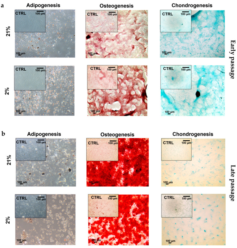
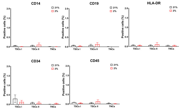
Tendon injuries caused by overuse or age-related deterioration are frequent. Incomplete knowledge of somatic tendon cell biology and their progenitors has hindered interventions for the effective repair of injured tendons. Here, we sought to compare and contrast distinct tendon-derived cell populations: type I and II tendon stem cells (TSCs) and tenocytes (TNCs). Porcine type I and II TSCs were isolated via the enzymatic digestion of distinct membranes (paratenon and endotenon, respectively), while tenocytes were isolated through an explant method. Resultant cell populations were characterized by morphology, differentiation, molecular, flow cytometry, and immunofluorescence analysis. Cells were isolated, cultured, and evaluated in two alternate oxygen concentrations (physiological (2%) and air (21%)) to determine the role of oxygen in cell biology determination within this relatively avascular tissue. The different cell populations demonstrated distinct proliferative potential, morphology, and transcript levels (both for tenogenic and stem cell markers). In contrast, all tendon-derived cell populations displayed multipotent differentiation potential and immunophenotypes (positive for CD90 and CD44). Type II TSCs emerged as the most promising tendon-derived cell population for expansion, given their enhanced proliferative potential, multipotency, and maintenance of a tenogenic profile at early and late passage. Moreover, in all cases, physoxia promoted the enhanced proliferation and maintenance of a tenogenic profile. These observations help shed light on the biological mechanisms of tendon cells, with the potential to aid in the development of novel therapeutic approaches for tendon disorders.

摘要

过度使用或与年龄相关的恶化导致的肌腱损伤很常见。对躯体肌腱细胞生物学及其祖细胞的不完全了解,阻碍了对受伤肌腱进行有效修复的干预措施。在这里,我们试图比较和对比不同的肌腱衍生细胞群体:I 型和 II 型肌腱干细胞(TSC)和肌腱细胞(TNC)。通过对不同膜(分别为腱旁组织和腱内膜)的酶消化,分离出猪 I 型和 II 型 TSC,而肌腱细胞则通过外植体方法分离。通过形态学、分化、分子、流式细胞术和免疫荧光分析对所得细胞群体进行了表征。将细胞在两种不同的氧浓度(生理浓度(2%)和空气浓度(21%))下进行分离、培养和评估,以确定氧气在这个相对无血管组织中对细胞生物学的作用。不同的细胞群体表现出不同的增殖潜力、形态和转录水平(包括肌腱和成体干细胞标志物)。相比之下,所有肌腱衍生细胞群体均显示出多能分化潜力和免疫表型(CD90 和 CD44 阳性)。考虑到 II 型 TSC 具有增强的增殖潜力、多能性以及在早期和晚期传代时保持肌腱表型,它是最有前途的肌腱衍生细胞群体,适合进行扩增。此外,在所有情况下,低氧条件都能促进增殖和保持肌腱表型。这些观察结果有助于阐明肌腱细胞的生物学机制,为肌腱疾病的新型治疗方法的发展提供了帮助。

https://cdn.ncbi.nlm.nih.gov/pmc/blobs/f018/10606148/c25dfee159b8/ijms-24-15107-g016.jpg
https://cdn.ncbi.nlm.nih.gov/pmc/blobs/f018/10606148/61761360bc5c/ijms-24-15107-g001.jpg
https://cdn.ncbi.nlm.nih.gov/pmc/blobs/f018/10606148/de21d98327aa/ijms-24-15107-g002.jpg
https://cdn.ncbi.nlm.nih.gov/pmc/blobs/f018/10606148/00b48ffef187/ijms-24-15107-g003.jpg
https://cdn.ncbi.nlm.nih.gov/pmc/blobs/f018/10606148/d553cf880477/ijms-24-15107-g004.jpg
https://cdn.ncbi.nlm.nih.gov/pmc/blobs/f018/10606148/1b49df96fb30/ijms-24-15107-g005.jpg
https://cdn.ncbi.nlm.nih.gov/pmc/blobs/f018/10606148/66d1b0855b37/ijms-24-15107-g006.jpg
https://cdn.ncbi.nlm.nih.gov/pmc/blobs/f018/10606148/4c2e21890751/ijms-24-15107-g007.jpg
https://cdn.ncbi.nlm.nih.gov/pmc/blobs/f018/10606148/9008a1a5b329/ijms-24-15107-g008.jpg
https://cdn.ncbi.nlm.nih.gov/pmc/blobs/f018/10606148/5f75cb4ba862/ijms-24-15107-g009.jpg
https://cdn.ncbi.nlm.nih.gov/pmc/blobs/f018/10606148/435cf262f594/ijms-24-15107-g010.jpg
https://cdn.ncbi.nlm.nih.gov/pmc/blobs/f018/10606148/e8f3090297bf/ijms-24-15107-g011.jpg
https://cdn.ncbi.nlm.nih.gov/pmc/blobs/f018/10606148/0321b352ed11/ijms-24-15107-g012.jpg
https://cdn.ncbi.nlm.nih.gov/pmc/blobs/f018/10606148/f239cccb0f2e/ijms-24-15107-g013.jpg
https://cdn.ncbi.nlm.nih.gov/pmc/blobs/f018/10606148/071cb0ebb748/ijms-24-15107-g014.jpg
https://cdn.ncbi.nlm.nih.gov/pmc/blobs/f018/10606148/f695b068e376/ijms-24-15107-g015.jpg
https://cdn.ncbi.nlm.nih.gov/pmc/blobs/f018/10606148/c25dfee159b8/ijms-24-15107-g016.jpg

相似文献

1
Endotenon-Derived Type II Tendon Stem Cells Have Enhanced Proliferative and Tenogenic Potential.腱内膜来源的 II 型肌腱干细胞具有增强的增殖和肌腱生成潜力。
Int J Mol Sci. 2023 Oct 12;24(20):15107. doi: 10.3390/ijms242015107.
2
Characterization of differential properties of rabbit tendon stem cells and tenocytes.兔肌腱干细胞和肌腱细胞差异特性的表征。
BMC Musculoskelet Disord. 2010 Jan 18;11:10. doi: 10.1186/1471-2474-11-10.
3
A comparison of the stem cell characteristics of murine tenocytes and tendon-derived stem cells.小鼠腱细胞与肌腱来源干细胞的干细胞特性比较。
BMC Musculoskelet Disord. 2018 Apr 12;19(1):116. doi: 10.1186/s12891-018-2038-2.
4
Tendon tissue engineering: Current progress towards an optimized tenogenic differentiation protocol for human stem cells.肌腱组织工程:当前在优化人干细胞的肌腱向分化方案方面的进展。
Acta Biomater. 2022 Jun;145:25-42. doi: 10.1016/j.actbio.2022.04.028. Epub 2022 Apr 22.
5
Effects of young extracellular matrix on the biological characteristics of aged tendon stem cells.年轻细胞外基质对老化肌腱干细胞生物学特性的影响。
Adv Clin Exp Med. 2018 Dec;27(12):1625-1630. doi: 10.17219/acem/75503.
6
Efficacy of tendon stem cells in fibroblast-derived matrix for tendon tissue engineering.肌腱干细胞在成纤维细胞衍生基质中对于肌腱组织工程的功效。
Cytotherapy. 2014 May;16(5):662-73. doi: 10.1016/j.jcyt.2013.07.014. Epub 2013 Oct 3.
7
Human Muscle-Derived Cells Are Capable of Tenogenic Differentiation and Contribution to Tendon Repair.人肌肉来源细胞能够进行肌腱分化并促进肌腱修复。
Am J Sports Med. 2023 Mar;51(3):786-797. doi: 10.1177/03635465221147486. Epub 2023 Feb 3.
8
Cystic fibrosis transmembrane conductance regulator mediates tenogenic differentiation of tendon-derived stem cells and tendon repair: accelerating tendon injury healing by intervening in its downstream signaling.囊性纤维化跨膜传导调节因子介导肌腱来源干细胞的成腱分化和肌腱修复:通过干预其下游信号通路加速肌腱损伤愈合
FASEB J. 2017 Sep;31(9):3800-3815. doi: 10.1096/fj.201601181R. Epub 2017 May 11.
9
CTGF induces tenogenic differentiation and proliferation of adipose-derived stromal cells.CTGF 诱导脂肪来源的基质细胞向肌腱细胞分化和增殖。
J Orthop Res. 2019 Mar;37(3):574-582. doi: 10.1002/jor.24248. Epub 2019 Feb 28.
10
EGR1 induces tenogenic differentiation of tendon stem cells and promotes rabbit rotator cuff repair.EGR1诱导肌腱干细胞向腱性分化并促进兔肩袖损伤修复。
Cell Physiol Biochem. 2015;35(2):699-709. doi: 10.1159/000369730. Epub 2015 Jan 28.

引用本文的文献

1
Tenogenic Cues Are Biochemically and Environmentally Distinct for Tendon Stem Cells and Mesenchymal/Stromal Stem Cells.对于肌腱干细胞和间充质/基质干细胞而言,成腱信号在生化和环境方面存在差异。
Stem Cells Int. 2025 May 13;2025:9047956. doi: 10.1155/sci/9047956. eCollection 2025.

本文引用的文献

1
Tendon tissue engineering: An overview of biologics to promote tendon healing and repair.肌腱组织工程:促进肌腱愈合与修复的生物制剂概述
J Tissue Eng. 2023 Sep 13;14:20417314231196275. doi: 10.1177/20417314231196275. eCollection 2023 Jan-Dec.
2
Fabrication of hydrogel microspheres microfluidics using inverse electron demand Diels-Alder click chemistry-based tetrazine-norbornene for drug delivery and cell encapsulation applications.基于逆电子需求 Diels-Alder 点击化学的四嗪-降冰片烯的水凝胶微球的微流控芯片的制备用于药物输送和细胞包封应用。
Biomater Sci. 2023 Jul 12;11(14):4972-4984. doi: 10.1039/d3bm00292f.
3
In Vitro Study of the Anti-inflammatory and Antifibrotic Activity of Tannic Acid-Coated Curcumin-Loaded Nanoparticles in Human Tenocytes.
鞣酸包覆姜黄素载纳米粒子在人腱细胞中的抗炎和抗纤维化活性的体外研究。
ACS Appl Mater Interfaces. 2023 May 17;15(19):23012-23023. doi: 10.1021/acsami.3c05322. Epub 2023 May 2.
4
Stem Cells from Healthy and Tendinopathic Human Tendons: Morphology, Collagen and Cytokines Expression and Their Response to T3 Thyroid Hormone.健康和病变人肌腱的干细胞:形态、胶原和细胞因子表达及其对 T3 甲状腺激素的反应。
Cells. 2022 Aug 16;11(16):2545. doi: 10.3390/cells11162545.
5
3D Biomimetic Scaffold for Growth Factor Controlled Delivery: An In-Vitro Study of Tenogenic Events on Wharton's Jelly Mesenchymal Stem Cells.用于生长因子控制释放的3D仿生支架:对沃顿胶间充质干细胞肌腱生成事件的体外研究
Pharmaceutics. 2021 Sep 10;13(9):1448. doi: 10.3390/pharmaceutics13091448.
6
Identification and Distinction of Tenocytes and Tendon-Derived Stem Cells.肌腱细胞与肌腱衍生干细胞的鉴定与区分
Front Cell Dev Biol. 2021 Apr 16;9:629515. doi: 10.3389/fcell.2021.629515. eCollection 2021.
7
Tendon Stem/Progenitor Cell Subpopulations and Their Implications in Tendon Biology.肌腱干/祖细胞亚群及其在肌腱生物学中的意义
Front Cell Dev Biol. 2021 Feb 18;9:631272. doi: 10.3389/fcell.2021.631272. eCollection 2021.
8
Characterization of the structure, vascularity, and stem/progenitor cell populations in porcine Achilles tendon (PAT).猪跟腱(PAT)的结构、血管和干细胞/祖细胞群体的特征。
Cell Tissue Res. 2021 May;384(2):367-387. doi: 10.1007/s00441-020-03379-3. Epub 2021 Jan 26.
9
In Vitro Innovation of Tendon Tissue Engineering Strategies.体外创新的肌腱组织工程策略。
Int J Mol Sci. 2020 Sep 14;21(18):6726. doi: 10.3390/ijms21186726.
10
Dose-Response Tendon-Specific Markers Induction by Growth Differentiation Factor-5 in Human Bone Marrow and Umbilical Cord Mesenchymal Stem Cells.生长分化因子 5 诱导人骨髓和脐带来源间充质干细胞产生肌腱特异性标志物。
Int J Mol Sci. 2020 Aug 17;21(16):5905. doi: 10.3390/ijms21165905.