Li Qi, Wang Yongli, Liu Huawen, Peng Hailang, Xiang Jianglin, Guo Shuliang
Department of Radiology, The First Affiliated Hospital of Chongqing Medical University, Chongqing, 400016, People's Republic of China.
Department of Infectious Disease, Chongqing University Three Gorges Hospital, Chongqing, 404000, People's Republic of China.
Infect Drug Resist. 2023 Oct 25;16:6795-6806. doi: 10.2147/IDR.S417062. eCollection 2023.
To investigate the computed tomography (CT) findings of SARs-CoV-2 Omicron variant in relation to respiratory viral loads determined by cycle threshold values in reverse-transcription polymerase chain reaction (RT-PCR).
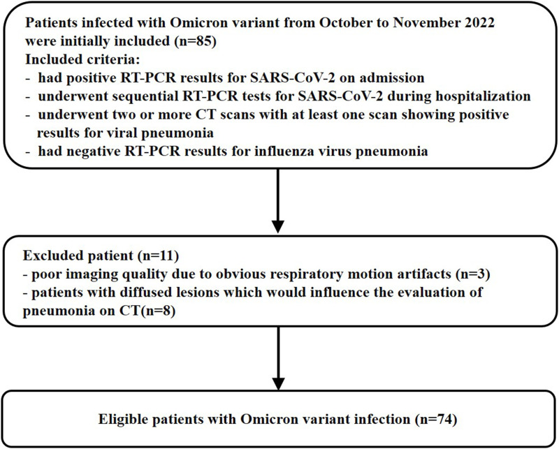
From October 2022 to November 2022, 74 hospitalized patients with Omicron were included in this retrospective study. The radiological features, CT involvement scores in relation to the respiratory viral load, and factors associated with imaging progression (IP) after the RT-PCR results turned negative were analyzed.
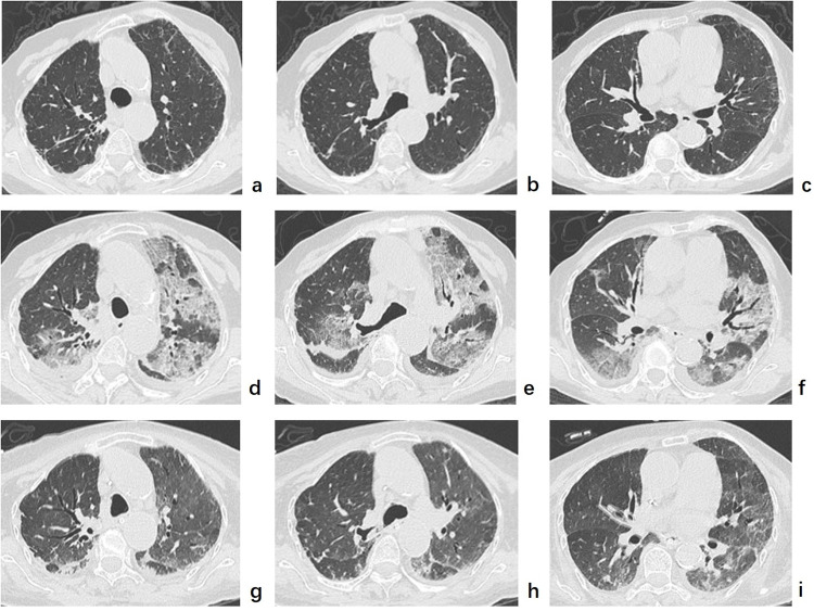
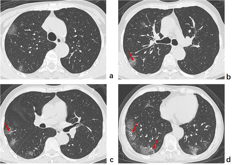
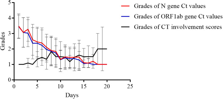
The most common CT patterns of Omicron were multiple round-like or patchy ground-glass opacity (GGO) or mixed GGO in the peripheral or diffuse areas. The grading of CT involvement scores exhibited an inverse pattern compared to viral loads from day 1 to day 8 and from day 13 to day 20 after diagnosis. Among the 65 patients with complete imaging data, 45 (69.23%) showed IP with clinical warning indicators of disease exacerbation negative in 34 and positive in 11. Patients with IP were older than those with non-IP (NIP); the erythrocyte sedimentation rates, procalcitonin levels, and D-dimer levels on admission of patients with IP were significantly higher than those of patients with NIP, whereas the immunoglobulin (Ig) G antibody level on admission and CT involvement score on initial CT of patients with IP were significantly lower than those of patients with NIP (all < 0.05).
For patients with Omicron, the IP of lung abnormalities is common when the viral load decreases. Under these circumstances, paying attention to clinical warming indicators of disease progression may contribute to better patient management and the mitigation of severe pneumonia.
探讨严重急性呼吸综合征冠状病毒2(SARS-CoV-2)奥密克戎变异株的计算机断层扫描(CT)表现与通过逆转录聚合酶链反应(RT-PCR)循环阈值确定的呼吸道病毒载量之间的关系。
2022年10月至2022年11月,74例住院的奥密克戎患者纳入本回顾性研究。分析了放射学特征、与呼吸道病毒载量相关的CT累及评分以及RT-PCR结果转阴后与影像进展(IP)相关的因素。
奥密克戎最常见的CT表现为外周或弥漫性区域的多发类圆形或斑片状磨玻璃影(GGO)或混合性GGO。诊断后第1天至第8天以及第13天至第20天,CT累及评分的分级与病毒载量呈相反模式。在65例有完整影像数据的患者中,45例(69.23%)出现IP,其中34例疾病加重的临床警示指标为阴性,11例为阳性。出现IP的患者比未出现IP(NIP)的患者年龄更大;出现IP的患者入院时的红细胞沉降率、降钙素原水平和D-二聚体水平显著高于NIP患者,而出现IP的患者入院时的免疫球蛋白(Ig)G抗体水平和初始CT的CT累及评分显著低于NIP患者(均P<0.05)。
对于奥密克戎患者,病毒载量下降时肺部异常的IP很常见。在这种情况下,关注疾病进展的临床警示指标可能有助于更好地管理患者并减轻重症肺炎。