Li Junping, Zhu Zhigang, Zhu Yuan, Li Jinqing, Li Kangbao, Zhong Weijie
Department of Geriatrics, Hematology & Oncology Ward, the Second Affiliated Hospital, School of Medicine, South China University of Technology, 510180, Guangzhou, Guangdong, China.
Department of Geriatrics, Gastroenterology Ward, the Second Affiliated Hospital, School of Medicine, South China University of Technology, 510180, Guangzhou, Guangdong, China.
Cell Death Discov. 2023 Nov 1;9(1):405. doi: 10.1038/s41420-023-01698-2.
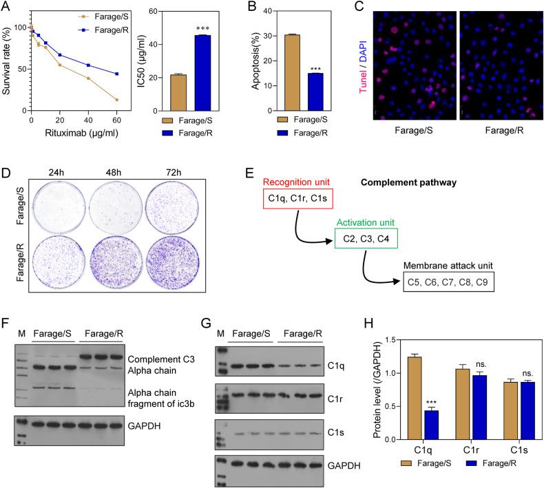
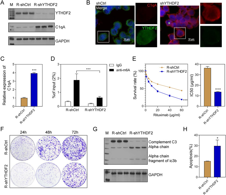
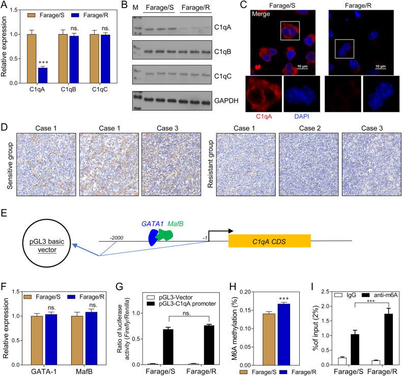
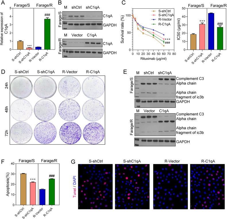
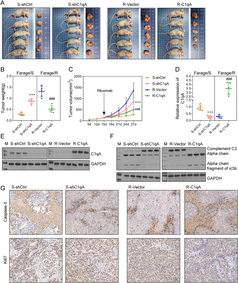
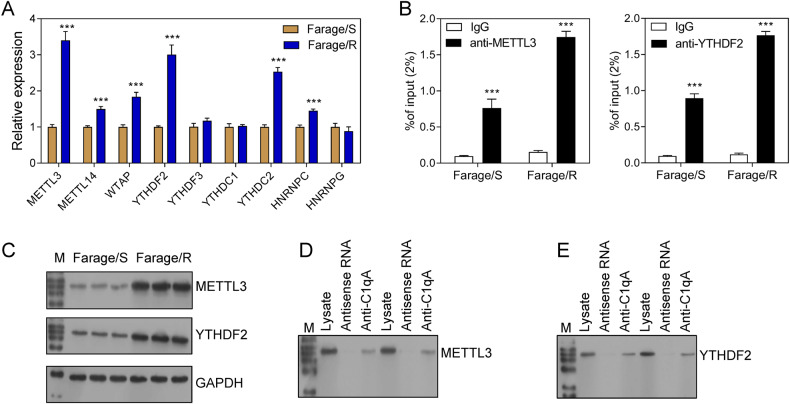
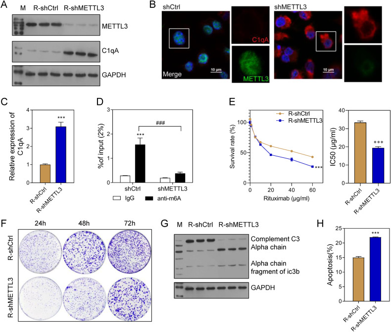
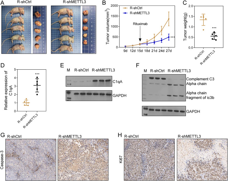
Rituximab has been incorporated into the standard treatment regimen for diffuse large B-cell lymphoma (DLBCL), and induces the death of tumor cells via complement-dependent cytotoxicity (CDC). Unfortunately, the resistance of DLBCL cells to Rituximab limits its clinical usefulness. It remains unclear whether the complement system is related to Rituximab resistance in DLBCL. A Rituximab-resistant DLBCL cell line (Farage/R) was generated under the stress of Rituximab. Constituent proteins of the complement system in wild-type Farage cells (Farage/S) and Farage/R cells were analyzed by qPCR, western blotting, and immunofluorescence. In vitro and in vivo knockdown and overexpression studies confirmed that the complement 1Q subcomponent A chain (C1qA) was a regulator of Rituximab resistance. Finally, the mechanism by which C1qA is regulated by mA methylation was explored. The reader and writer were identified by pull-down studies and RIP-qPCR. Activity of the complement system in Farage/R cells was suppressed. C1qA expression was reduced in Farage/R cells due to post-transcriptional regulation. Furthermore, in vitro and in vivo results showed that C1qA knockdown in Farage/S cells decreased their sensitivity to Rituximab, and C1qA overexpression in Farage/R cells attenuated the Rituximab resistance of those cells. Moreover, METTL3 and YTHDF2 were proven to be the reader and writer for mA methylation of C1qA, respectively. Knockdown of METTL3 or YTHDF2 in Farage/R cells up-regulated C1qA expression and reduced their resistance to Rituximab. In summary, the aberrant downregulation of C1qA was related to Rituximab resistance in DLBCL cells, and C1qA was found to be regulated by METTL3- and YTHDF2-mediated m6A methylation. Enhancing the response of the complement system via regulation of C1qA might be an effective strategy for inhibiting Rituximab resistance in DLBCL.
利妥昔单抗已被纳入弥漫性大B细胞淋巴瘤(DLBCL)的标准治疗方案,并通过补体依赖性细胞毒性(CDC)诱导肿瘤细胞死亡。不幸的是,DLBCL细胞对利妥昔单抗的耐药性限制了其临床应用。目前尚不清楚补体系统是否与DLBCL中利妥昔单抗耐药性有关。在利妥昔单抗的压力下产生了一种利妥昔单抗耐药的DLBCL细胞系(Farage/R)。通过qPCR、蛋白质免疫印迹和免疫荧光分析野生型Farage细胞(Farage/S)和Farage/R细胞中补体系统的组成蛋白。体外和体内的敲低和过表达研究证实,补体1Q亚成分A链(C1qA)是利妥昔单抗耐药性的调节因子。最后,探讨了C1qA受mA甲基化调节的机制。通过下拉实验和RIP-qPCR鉴定了读取器和写入器。Farage/R细胞中补体系统的活性受到抑制。由于转录后调控,Farage/R细胞中C1qA表达降低。此外,体外和体内结果表明,Farage/S细胞中C1qA敲低降低了它们对利妥昔单抗的敏感性,而Farage/R细胞中C1qA过表达减弱了这些细胞对利妥昔单抗的耐药性。此外,METTL3和YTHDF2分别被证明是C1qA的mA甲基化的读取器和写入器。Farage/R细胞中METTL3或YTHDF2敲低上调了C1qA表达并降低了它们对利妥昔单抗的耐药性。总之,C1qA的异常下调与DLBCL细胞中利妥昔单抗耐药性有关,并且发现C1qA受METTL3和YTHDF2介导的m6A甲基化调节。通过调节C1qA增强补体系统的反应可能是抑制DLBCL中利妥昔单抗耐药性的有效策略。