Marsh Donald J, Wexler Anthony S, Holstein-Rathlou Niels-Henrik
Department of Medical Sciences, Division of Medicine and Biological Sciences, Brown University, Providence, RI, United States.
Departments of Biomedical Engineering, and Mechanical and Aerospace Engineering, University of California Davis, Davis, CA, United States.
Front Netw Physiol. 2023 Oct 19;3:1254964. doi: 10.3389/fnetp.2023.1254964. eCollection 2023.
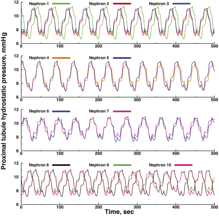
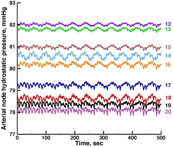
Blood flow and glomerular filtration in the kidney are regulated by two mechanisms acting on the afferent arteriole of each nephron. The two mechanisms operate as limit cycle oscillators, each responding to a different signal. The myogenic mechanism is sensitive to a transmural pressure difference across the wall of the arteriole, and tubuloglomerular feedback (TGF) responds to the NaCl concentration in tubular fluid flowing into the nephron's distal tubule,. The two mechanisms interact with each other, synchronize, cause oscillations in tubular flow and pressure, and form a bimodal electrical signal that propagates into the arterial network. The electrical signal enables nephrons adjacent to each other in the arterial network to synchronize, but non-adjacent nephrons do not synchronize. The arteries supplying the nephrons have the morphologic characteristics of a rooted tree network, with 3 motifs characterizing nephron distribution. We developed a model of 10 nephrons and their afferent arterioles in an arterial network that reproduced these structural characteristics, with half of its components on the renal surface, where experimental data suitable for model validation is available, and the other half below the surface, from which no experimental data has been reported. The model simulated several interactions: TGF-myogenic in each nephron with TGF modulating amplitude and frequency of the myogenic oscillation; adjacent nephron-nephron with strong coupling; non-adjacent nephron-nephron, with weak coupling because of electrical signal transmission through electrically conductive arterial walls; and coupling involving arterial nodal pressure at the ends of each arterial segment, and between arterial nodes and the afferent arterioles originating at the nodes. The model predicted full synchronization between adjacent nephrons pairs and partial synchronization among weakly coupled nephrons, reproducing experimental findings. The model also predicted aperiodic fluctuations of tubular and arterial pressures lasting longer than TGF oscillations in nephrons, again confirming experimental observations. The model did not predict complete synchronization of all nephrons.
肾脏中的血流和肾小球滤过由作用于每个肾单位入球小动脉的两种机制调节。这两种机制作为极限环振荡器运行,各自对不同的信号作出反应。肌源机制对小动脉壁上的跨壁压差敏感,而管球反馈(TGF)则对流入肾单位远端小管的小管液中的NaCl浓度作出反应。这两种机制相互作用、同步,引起小管流量和压力的振荡,并形成一种双峰电信号,该信号传播到动脉网络中。该电信号使动脉网络中相邻的肾单位能够同步,但不相邻的肾单位则不能同步。供应肾单位的动脉具有根树状网络的形态特征,有3种基序表征肾单位的分布。我们建立了一个包含10个肾单位及其在动脉网络中的入球小动脉的模型,该模型再现了这些结构特征,其一半组件位于肾表面,在那里有适合模型验证的实验数据,另一半在表面以下,尚未有该部位的实验数据报道。该模型模拟了几种相互作用:每个肾单位中的TGF-肌源相互作用,其中TGF调节肌源振荡的幅度和频率;相邻肾单位-肾单位之间具有强耦合;不相邻肾单位-肾单位之间,由于电信号通过导电动脉壁传输而具有弱耦合;以及涉及每个动脉段末端的动脉节点压力,以及动脉节点与起源于这些节点的入球小动脉之间的耦合。该模型预测相邻肾单位对之间会完全同步,弱耦合肾单位之间会部分同步,再现了实验结果。该模型还预测了小管和动脉压力的非周期性波动,其持续时间比肾单位中的TGF振荡更长,再次证实了实验观察结果。该模型没有预测所有肾单位会完全同步。