Department of Oncology, The Second Affiliated Hospital of Anhui Medical University, No. 678 Fu Rong Road, Hefei, 230601, China.
Department of Oncology, Anhui Medical University, Hefei, 230000, China.
BMC Cancer. 2023 Nov 10;23(1):1095. doi: 10.1186/s12885-023-11620-9.
The aim of this study was to investigate the influence of serum iron levels in advanced gastric cancer (GC) patients treated with programmed cell death protein-1 (PD-1) inhibitors.
We retrospectively reviewed 149 GC patients who were treated with PD-1 inhibitors at our center. Clinicopathological characteristics, laboratory data, and clinical outcomes were analyzed.
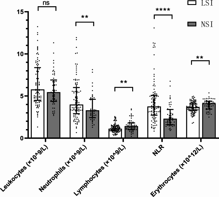
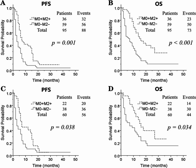
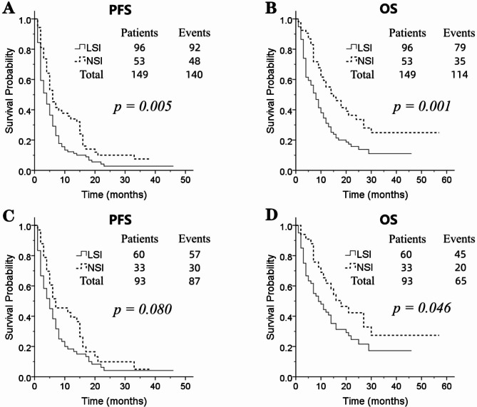
Multivariate analysis showed that Eastern Cooperative Oncology Group performance status (ECOG PS), histological subtype, and baseline serum iron levels were independent prognostic factors for overall survival (OS), while ECOG PS, multiple metastatic sites, and baseline serum iron levels were independent prognostic factors for progression-free survival (PFS). Patients with baseline low serum iron levels (LSI) had a significantly shorter median OS and PFS compared to patients with normal serum iron levels (NSI) (Median OS: 7 vs. 14 months, p = 0.001; median PFS: 3 vs. 5 months, p = 0.005). Patients with baseline LSI had a disease control rate (DCR) of 58.3% at 2 months after PD-1 inhibitor initiation (M2), compared to 81.1% in patients with NSI (p = 0.005). Patients with baseline LSI had a DCR of 43.8% at 4 months, compared to 64.2% in patients with NSI (p = 0.017).
LSI was associated with worse OS, PFS, and DCR in GC patients treated with PD-1 inhibitors and might be a quick and efficient biomarker to predict the efficacy of PD-1 inhibitors.
本研究旨在探讨程序性死亡蛋白-1(PD-1)抑制剂治疗晚期胃癌(GC)患者时血清铁水平的影响。
我们回顾性分析了在我院接受 PD-1 抑制剂治疗的 149 例 GC 患者。分析了临床病理特征、实验室数据和临床结局。
多因素分析显示,东部肿瘤协作组体力状态(ECOG PS)、组织学亚型和基线血清铁水平是总生存期(OS)的独立预后因素,而 ECOG PS、多个转移部位和基线血清铁水平是无进展生存期(PFS)的独立预后因素。基线低血清铁水平(LSI)患者的中位 OS 和 PFS 明显短于正常血清铁水平(NSI)患者(中位 OS:7 个月比 14 个月,p=0.001;中位 PFS:3 个月比 5 个月,p=0.005)。PD-1 抑制剂治疗 2 个月时(M2),基线 LSI 患者的疾病控制率(DCR)为 58.3%,而 NSI 患者的 DCR 为 81.1%(p=0.005)。基线 LSI 患者在 4 个月时的 DCR 为 43.8%,而 NSI 患者的 DCR 为 64.2%(p=0.017)。
LSI 与 GC 患者接受 PD-1 抑制剂治疗后的 OS、PFS 和 DCR 较差相关,可能是一种快速有效的预测 PD-1 抑制剂疗效的生物标志物。