Department of Oral and Cranio-maxillofacial Surgery, Shanghai Ninth People's Hospital, College of Stomatology, Shanghai Jiao Tong University School of Medicine, Shanghai, 200011, China.
National Center for Stomatology & National Clinical Research Center for Oral Diseases, Shanghai, 200011, China.
BMC Oral Health. 2023 Nov 17;23(1):876. doi: 10.1186/s12903-023-03446-5.
Accurate cephalometric analysis plays a vital role in the diagnosis and subsequent surgical planning in orthognathic and orthodontics treatment. However, manual digitization of anatomical landmarks in computed tomography (CT) is subject to limitations such as low accuracy, poor repeatability and excessive time consumption. Furthermore, the detection of landmarks has more difficulties on individuals with dentomaxillofacial deformities than normal individuals. Therefore, this study aims to develop a deep learning model to automatically detect landmarks in CT images of patients with dentomaxillofacial deformities.
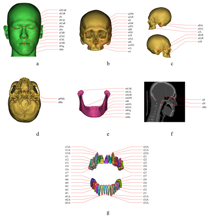
Craniomaxillofacial (CMF) CT data of 80 patients with dentomaxillofacial deformities were collected for model development. 77 anatomical landmarks digitized by experienced CMF surgeons in each CT image were set as the ground truth. 3D UX-Net, the cutting-edge medical image segmentation network, was adopted as the backbone of model architecture. Moreover, a new region division pattern for CMF structures was designed as a training strategy to optimize the utilization of computational resources and image resolution. To evaluate the performance of this model, several experiments were conducted to make comparison between the model and manual digitization approach.
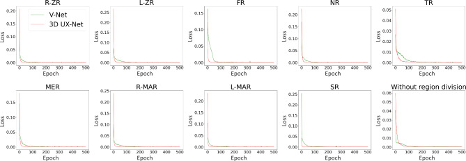
The training set and the validation set included 58 and 22 samples respectively. The developed model can accurately detect 77 landmarks on bone, soft tissue and teeth with a mean error of 1.81 ± 0.89 mm. Removal of region division before training significantly increased the error of prediction (2.34 ± 1.01 mm). In terms of manual digitization, the inter-observer and intra-observer variations were 1.27 ± 0.70 mm and 1.01 ± 0.74 mm respectively. In all divided regions except Teeth Region (TR), our model demonstrated equivalent performance to experienced CMF surgeons in landmarks detection (p > 0.05).
The developed model demonstrated excellent performance in detecting craniomaxillofacial landmarks when considering manual digitization work of expertise as benchmark. It is also verified that the region division pattern designed in this study remarkably improved the detection accuracy.
准确的头影测量分析在正颌和正畸治疗的诊断和后续手术计划中起着至关重要的作用。然而,在计算机断层扫描(CT)中手动数字化解剖标志存在准确性低、可重复性差和耗时过多等局限性。此外,与正常个体相比,牙颌面畸形患者的标志检测更加困难。因此,本研究旨在开发一种深度学习模型,以自动检测牙颌面畸形患者 CT 图像中的标志。
收集 80 例牙颌面畸形患者的颅颌面(CMF)CT 数据,用于模型开发。在每张 CT 图像中,由经验丰富的 CMF 外科医生数字化 77 个解剖标志,并将其作为ground truth。采用最先进的医学图像分割网络 3D UX-Net 作为模型架构的骨干。此外,还设计了一种新的 CMF 结构区域划分模式作为训练策略,以优化计算资源和图像分辨率的利用。为了评估该模型的性能,进行了多项实验,将模型与手动数字化方法进行了比较。
训练集和验证集分别包含 58 个和 22 个样本。开发的模型可以准确地检测到骨骼、软组织和牙齿上的 77 个标志,平均误差为 1.81±0.89mm。在训练前去除区域划分会显著增加预测误差(2.34±1.01mm)。在手动数字化方面,观察者间和观察者内的变异分别为 1.27±0.70mm 和 1.01±0.74mm。在除牙齿区域(TR)之外的所有划分区域中,我们的模型在标志检测方面表现出与经验丰富的 CMF 外科医生相当的性能(p>0.05)。
当将手动数字化工作的专业知识作为基准时,开发的模型在检测颅颌面标志方面表现出优异的性能。还验证了本研究中设计的区域划分模式显著提高了检测精度。