• 文献检索
  • 文档翻译
  • 深度研究
  • 学术资讯
  • Suppr Zotero 插件Zotero 插件
  • 邀请有礼
  • 套餐&价格
  • 历史记录
应用&插件
Suppr Zotero 插件Zotero 插件浏览器插件Mac 客户端Windows 客户端微信小程序
定价
高级版会员购买积分包购买API积分包
服务
文献检索文档翻译深度研究API 文档MCP 服务
关于我们
关于 Suppr公司介绍联系我们用户协议隐私条款
关注我们

Suppr 超能文献

核心技术专利:CN118964589B侵权必究
粤ICP备2023148730 号-1Suppr @ 2026

文献检索

告别复杂PubMed语法,用中文像聊天一样搜索,搜遍4000万医学文献。AI智能推荐,让科研检索更轻松。

立即免费搜索

文件翻译

保留排版,准确专业,支持PDF/Word/PPT等文件格式,支持 12+语言互译。

免费翻译文档

深度研究

AI帮你快速写综述,25分钟生成高质量综述,智能提取关键信息,辅助科研写作。

立即免费体验

不同真菌植物病原体中II类疏水蛋白家族的计算机模拟评估、系统发育分析和结构建模

In Silico Evaluation, Phylogenetic Analysis, and Structural Modeling of the Class II Hydrophobin Family from Different Fungal Phytopathogens.

作者信息

Bouqellah Nahla A, Farag Peter F

机构信息

Department of Biology, College of Science, Taibah University, P.O. Box 344, Al Madinah Al Munawwarah 42317-8599, Saudi Arabia.

Department of Microbiology, Faculty of Science, Ain Shams University, Cairo 11566, Egypt.

出版信息

Microorganisms. 2023 Oct 26;11(11):2632. doi: 10.3390/microorganisms11112632.

DOI:10.3390/microorganisms11112632
PMID:38004644
原文链接:https://pmc.ncbi.nlm.nih.gov/articles/PMC10672791/
Abstract

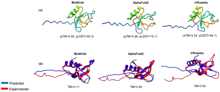
The class II hydrophobin group (HFBII) is an extracellular group of proteins that contain the HFBII domain and eight conserved cysteine residues. These proteins are exclusively secreted by fungi and have multiple functions with a probable role as effectors. In the present study, a total of 45 amino acid sequences of hydrophobin class II proteins from different phytopathogenic fungi were retrieved from the NCBI database. We used the integration of well-designed bioinformatic tools to characterize and predict their physicochemical parameters, novel motifs, 3D structures, multiple sequence alignment (MSA), evolution, and functions as effector proteins through molecular docking. The results revealed new features for these protein members. The ProtParam tool detected the hydrophobicity properties of all proteins except for one hydrophilic protein (KAI3335996.1). Out of 45 proteins, six of them were detected as GPI-anchored proteins by the PredGPI server. Different 3D structure templates with high pTM scores were designed by Multifold v1, AlphaFold2, and trRosetta. Most of the studied proteins were anticipated as apoplastic effectors and matched with the ghyd5 gene of as virulence factors. A protein-protein interaction (PPI) analysis unraveled the molecular function of this group as GTP-binding proteins, while a molecular docking analysis detected a chitin-binding effector role. From the MSA analysis, it was observed that the HFBII sequences shared conserved 2 Pro (P) and 2 Gly (G) amino acids besides the known eight conserved cysteine residues. The evolutionary analysis and phylogenetic tree provided evidence of episodic diversifying selection at the branch level using the aBSREL tool. A detailed in silico analysis of this family and the present findings will provide a better understanding of the HFBII characters and evolutionary relationships, which could be very useful in future studies.

摘要

II类疏水蛋白家族(HFBII)是一类细胞外蛋白,包含HFBII结构域和八个保守的半胱氨酸残基。这些蛋白仅由真菌分泌,具有多种功能,可能作为效应子发挥作用。在本研究中,从NCBI数据库中检索了来自不同植物病原真菌的45个II类疏水蛋白的氨基酸序列。我们使用精心设计的生物信息学工具进行整合,以表征和预测它们的物理化学参数、新基序、三维结构、多序列比对(MSA)、进化以及通过分子对接作为效应蛋白的功能。结果揭示了这些蛋白成员的新特征。ProtParam工具检测到除一种亲水性蛋白(KAI3335996.1)外所有蛋白的疏水性。在45种蛋白中,PredGPI服务器检测到其中六种为糖基磷脂酰肌醇(GPI)锚定蛋白。通过Multifold v1、AlphaFold2和trRosetta设计了具有高pTM分数的不同三维结构模板。大多数研究的蛋白被预测为质外体效应子,并与作为毒力因子的ghyd5基因匹配。蛋白质-蛋白质相互作用(PPI)分析揭示了该家族作为GTP结合蛋白的分子功能,而分子对接分析检测到其几丁质结合效应子作用。从MSA分析中观察到,除了已知的八个保守半胱氨酸残基外,HFBII序列还共享保守的2个脯氨酸(P)和2个甘氨酸(G)氨基酸。进化分析和系统发育树使用aBSREL工具在分支水平上提供了间歇性多样化选择的证据。对该家族进行详细的计算机分析以及本研究结果将有助于更好地理解HFBII的特征和进化关系,这在未来的研究中可能非常有用。

https://cdn.ncbi.nlm.nih.gov/pmc/blobs/22ea/10672791/2f81f11ae64c/microorganisms-11-02632-g013.jpg
https://cdn.ncbi.nlm.nih.gov/pmc/blobs/22ea/10672791/dc6efd6a8969/microorganisms-11-02632-g001.jpg
https://cdn.ncbi.nlm.nih.gov/pmc/blobs/22ea/10672791/9e6bfaec3af4/microorganisms-11-02632-g002.jpg
https://cdn.ncbi.nlm.nih.gov/pmc/blobs/22ea/10672791/0c071ed80f6f/microorganisms-11-02632-g003.jpg
https://cdn.ncbi.nlm.nih.gov/pmc/blobs/22ea/10672791/12ba1689154a/microorganisms-11-02632-g004.jpg
https://cdn.ncbi.nlm.nih.gov/pmc/blobs/22ea/10672791/7103ea42f904/microorganisms-11-02632-g005.jpg
https://cdn.ncbi.nlm.nih.gov/pmc/blobs/22ea/10672791/b6c3a963cfcd/microorganisms-11-02632-g006.jpg
https://cdn.ncbi.nlm.nih.gov/pmc/blobs/22ea/10672791/7808a432e1c3/microorganisms-11-02632-g007.jpg
https://cdn.ncbi.nlm.nih.gov/pmc/blobs/22ea/10672791/0b32e1badb7e/microorganisms-11-02632-g008.jpg
https://cdn.ncbi.nlm.nih.gov/pmc/blobs/22ea/10672791/9908ec2c4267/microorganisms-11-02632-g009.jpg
https://cdn.ncbi.nlm.nih.gov/pmc/blobs/22ea/10672791/f49f0adf82ad/microorganisms-11-02632-g010.jpg
https://cdn.ncbi.nlm.nih.gov/pmc/blobs/22ea/10672791/781968184359/microorganisms-11-02632-g011.jpg
https://cdn.ncbi.nlm.nih.gov/pmc/blobs/22ea/10672791/b0e2596b3d41/microorganisms-11-02632-g012.jpg
https://cdn.ncbi.nlm.nih.gov/pmc/blobs/22ea/10672791/2f81f11ae64c/microorganisms-11-02632-g013.jpg

相似文献

1
In Silico Evaluation, Phylogenetic Analysis, and Structural Modeling of the Class II Hydrophobin Family from Different Fungal Phytopathogens.不同真菌植物病原体中II类疏水蛋白家族的计算机模拟评估、系统发育分析和结构建模
Microorganisms. 2023 Oct 26;11(11):2632. doi: 10.3390/microorganisms11112632.
2
Investigation of the role hydrophobin monomer loops using hybrid models via molecular dynamics simulation.利用分子动力学模拟的混合模型研究疏水单体环的作用。
Colloids Surf B Biointerfaces. 2019 Jan 1;173:128-138. doi: 10.1016/j.colsurfb.2018.09.062. Epub 2018 Sep 26.
3
Cysteine-Rich Hydrophobin Gene Family: Genome Wide Analysis, Phylogeny and Transcript Profiling in .富含半胱氨酸的疏水蛋白基因家族:全基因组分析、系统发育及转录谱分析
Int J Mol Sci. 2021 Jan 11;22(2):643. doi: 10.3390/ijms22020643.
4
Predicting the self-assembly film structure of class II hydrophobin NC2 and estimating its structural characteristics.预测 II 类疏水蛋白 NC2 的自组装膜结构并估计其结构特征。
Colloids Surf B Biointerfaces. 2020 Nov;195:111269. doi: 10.1016/j.colsurfb.2020.111269. Epub 2020 Jul 26.
5
Identification of new members of hydrophobin family using primary structure analysis.利用一级结构分析鉴定疏水蛋白家族的新成员。
BMC Bioinformatics. 2006 Dec 12;7 Suppl 4(Suppl 4):S16. doi: 10.1186/1471-2105-7-S4-S16.
6
Atomic resolution structure of the HFBII hydrophobin, a self-assembling amphiphile.HFBII疏水蛋白的原子分辨率结构,一种自组装两亲分子。
J Biol Chem. 2004 Jan 2;279(1):534-9. doi: 10.1074/jbc.M309650200. Epub 2003 Oct 10.
7
Interaction and comparison of a class I hydrophobin from Schizophyllum commune and class II hydrophobins from Trichoderma reesei.裂褶菌的I类疏水蛋白与里氏木霉的II类疏水蛋白的相互作用及比较
Biomacromolecules. 2006 Apr;7(4):1295-301. doi: 10.1021/bm050676s.
8
Lack of evidence for a role of hydrophobins in conferring surface hydrophobicity to conidia and hyphae of Botrytis cinerea.缺乏证据表明亲脂蛋白在赋予灰葡萄孢分生孢子和菌丝表面疏水性方面的作用。
BMC Microbiol. 2011 Jan 13;11:10. doi: 10.1186/1471-2180-11-10.
9
Molecular dynamics simulations of the hydrophobin SC3 at a hydrophobic/hydrophilic interface.疏水蛋白SC3在疏水/亲水界面的分子动力学模拟。
Proteins. 2006 Sep 1;64(4):863-73. doi: 10.1002/prot.20936.
10
Evolutionary analysis of hydrophobin gene family in two wood-degrading basidiomycetes, Phlebia brevispora and Heterobasidion annosum s.l.两株木质腐朽担子菌(栓菌和粗皮侧耳)中疏水蛋白基因家族的进化分析
BMC Evol Biol. 2013 Nov 4;13:240. doi: 10.1186/1471-2148-13-240.

引用本文的文献

1
AlphaFold modeling uncovers global structural features of class I and class II fungal hydrophobins.AlphaFold建模揭示了I类和II类真菌疏水蛋白的整体结构特征。
Protein Sci. 2025 Sep;34(9):e70279. doi: 10.1002/pro.70279.
2
Integrative workflows for the characterization of hydrophobin and cerato-platanin in the marine fungus Paradendryphiella salina.盐生拟青霉中疏水性蛋白和角蛋白- platanin 的特性分析的综合工作流程。
Arch Microbiol. 2024 Aug 23;206(9):385. doi: 10.1007/s00203-024-04087-0.

本文引用的文献

1
Identification and Expression Analysis of the Alfin-like Gene Family in Tomato and the Role of in Salt and Drought Stresses.番茄中Alfin样基因家族的鉴定与表达分析及其在盐胁迫和干旱胁迫中的作用
Plants (Basel). 2023 Jul 31;12(15):2829. doi: 10.3390/plants12152829.
2
Secretome Analysis for a New Strain of the Blackleg Fungus Reveals Candidate Proteins for Effectors and Virulence Factors.黑腿病菌新菌株的分泌蛋白质组分析揭示了效应蛋白和毒力因子的候选蛋白。
J Fungi (Basel). 2023 Jul 11;9(7):740. doi: 10.3390/jof9070740.
3
Insights on Novel Effectors and Characterization of Metacaspase (RS107_6) as a Potential Cell Death-Inducing Protein in .
关于新型效应子的见解以及对metacaspase(RS107_6)作为[具体物种]中一种潜在的诱导细胞死亡蛋白的表征
Microorganisms. 2023 Apr 1;11(4):920. doi: 10.3390/microorganisms11040920.
4
Prediction of protein structures, functions and interactions using the IntFOLD7, MultiFOLD and ModFOLDdock servers.使用 IntFOLD7、MultiFOLD 和 ModFOLDdock 服务器预测蛋白质结构、功能和相互作用。
Nucleic Acids Res. 2023 Jul 5;51(W1):W274-W280. doi: 10.1093/nar/gkad297.
5
In Silico Characterization of the Secretome of the Fungal Pathogen , the Causal Agent of Date Palm Black Scorch Disease.枣椰树黑焦病病原体——一种真菌病原体的分泌蛋白组的计算机模拟特征分析
J Fungi (Basel). 2023 Feb 27;9(3):303. doi: 10.3390/jof9030303.
6
Plant Defense Elicitation by the Hydrophobin Cerato-Ulmin and Correlation with Its Structural Features.几丁质-乌来糖蛋白通过疏水性诱导植物防御及其与结构特征的相关性。
Int J Mol Sci. 2023 Jan 23;24(3):2251. doi: 10.3390/ijms24032251.
7
Using evolutionary data to make sense of macromolecules with a "face-lifted" ConSurf.利用进化数据,通过“改头换面”的 ConSurf 来理解大分子。
Protein Sci. 2023 Mar;32(3):e4582. doi: 10.1002/pro.4582.
8
Molecular Evolution of the Bactericidal/Permeability-Increasing Protein (BPIFA1) Regulating the Innate Immune Responses in Mammals.杀菌/通透性增加蛋白(BPIFA1)在哺乳动物固有免疫反应中的分子进化调控
Genes (Basel). 2022 Dec 21;14(1):15. doi: 10.3390/genes14010015.
9
Prediction of effector protein structures from fungal phytopathogens enables evolutionary analyses.从真菌植物病原体中预测效应蛋白结构可进行进化分析。
Nat Microbiol. 2023 Jan;8(1):174-187. doi: 10.1038/s41564-022-01287-6. Epub 2023 Jan 5.
10
The STRING database in 2023: protein-protein association networks and functional enrichment analyses for any sequenced genome of interest.2023 年的 STRING 数据库:针对任何感兴趣的测序基因组的蛋白质-蛋白质关联网络和功能富集分析。
Nucleic Acids Res. 2023 Jan 6;51(D1):D638-D646. doi: 10.1093/nar/gkac1000.