He Sheng, Guan Yi, Cheng Chia Hsin, Moore Tara L, Luebke Jennifer I, Killiany Ronald J, Rosene Douglas L, Koo Bang-Bon, Ou Yangming
Harvard Medical School, Boston Children's Hospital, Boston, MA, United States.
Department of Anatomy & Neurobiology, Boston University Chobanian and Avedisian School of Medicine, Boston, MA, United States.
Front Aging Neurosci. 2023 Nov 1;15:1249415. doi: 10.3389/fnagi.2023.1249415. eCollection 2023.
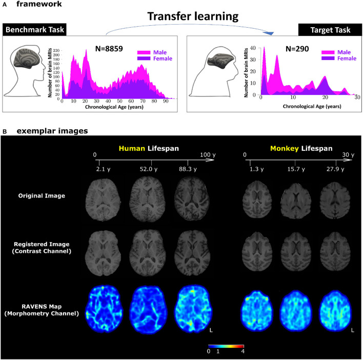
The application of artificial intelligence (AI) to summarize a whole-brain magnetic resonance image (MRI) into an effective "brain age" metric can provide a holistic, individualized, and objective view of how the brain interacts with various factors (e.g., genetics and lifestyle) during aging. Brain age predictions using deep learning (DL) have been widely used to quantify the developmental status of human brains, but their wider application to serve biomedical purposes is under criticism for requiring large samples and complicated interpretability. Animal models, i.e., rhesus monkeys, have offered a unique lens to understand the human brain - being a species in which aging patterns are similar, for which environmental and lifestyle factors are more readily controlled. However, applying DL methods in animal models suffers from data insufficiency as the availability of animal brain MRIs is limited compared to many thousands of human MRIs. We showed that transfer learning can mitigate the sample size problem, where transferring the pre-trained AI models from 8,859 human brain MRIs improved monkey brain age estimation accuracy and stability. The highest accuracy and stability occurred when transferring the 3D ResNet [mean absolute error (MAE) = 1.83 years] and the 2D global-local transformer (MAE = 1.92 years) models. Our models identified the frontal white matter as the most important feature for monkey brain age predictions, which is consistent with previous histological findings. This first DL-based, anatomically interpretable, and adaptive brain age estimator could broaden the application of AI techniques to various animal or disease samples and widen opportunities for research in non-human primate brains across the lifespan.
将人工智能(AI)应用于将全脑磁共振成像(MRI)总结为有效的“脑龄”指标,可以提供关于大脑在衰老过程中如何与各种因素(如基因和生活方式)相互作用的整体、个性化和客观的观点。使用深度学习(DL)进行脑龄预测已被广泛用于量化人类大脑的发育状态,但其在生物医学目的方面的更广泛应用因需要大量样本和复杂的可解释性而受到批评。动物模型,即恒河猴,为理解人类大脑提供了一个独特的视角——作为一种衰老模式相似的物种,其环境和生活方式因素更容易控制。然而,在动物模型中应用DL方法存在数据不足的问题,因为与数千个人类MRI相比,动物脑MRI的可用性有限。我们表明,迁移学习可以缓解样本量问题,即将预训练的AI模型从8859个人类脑MRI迁移过来,可以提高猴脑年龄估计的准确性和稳定性。当迁移3D ResNet(平均绝对误差[MAE]=1.83岁)和2D全局-局部变压器(MAE=1.92岁)模型时,准确性和稳定性最高。我们的模型确定额叶白质是猴脑年龄预测中最重要的特征,这与先前的组织学研究结果一致。这种首个基于DL的、具有解剖学可解释性的自适应脑龄估计器可以将AI技术的应用扩展到各种动物或疾病样本,并为整个生命周期内非人类灵长类动物大脑的研究提供更多机会。