Szelenyi Eric R, Navarrete Jovana S, Murry Alexandria D, Zhang Yizhe, Girven Kasey S, Kuo Lauren, Cline Marcella M, Bernstein Mollie X, Burdyniuk Mariia, Bowler Bryce, Goodwin Nastacia L, Juarez Barbara, Zweifel Larry S, Golden Sam A
University of Washington Center of Excellence in Neurobiology of Addiction, Pain, and Emotion (NAPE), Seattle, WA, USA.
University of Washington, Department of Biological Structure, Seattle, WA, USA.
bioRxiv. 2023 Dec 12:2023.11.22.568319. doi: 10.1101/2023.11.22.568319.
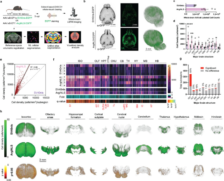
High-throughput volumetric fluorescent microscopy pipelines can spatially integrate whole-brain structure and function at the foundational level of single-cells. However, conventional fluorescent protein (FP) modifications used to discriminate single-cells possess limited efficacy or are detrimental to cellular health. Here, we introduce a synthetic and non-deleterious nuclear localization signal (NLS) tag strategy, called 'Arginine-rich NLS' (ArgiNLS), that optimizes genetic labeling and downstream image segmentation of single-cells by restricting FP localization near-exclusively in the nucleus through a poly-arginine mechanism. A single N-terminal ArgiNLS tag provides modular nuclear restriction consistently across spectrally separate FP variants. ArgiNLS performance in vivo displays functional conservation across major cortical cell classes, and in response to both local and systemic brain wide AAV administration. Crucially, the high signal-to-noise ratio afforded by ArgiNLS enhances ML-automated segmentation of single-cells due to rapid classifier training and enrichment of labeled cell detection within 2D brain sections or 3D volumetric whole-brain image datasets, derived from both staining-amplified and native signal. This genetic strategy provides a simple and flexible basis for precise image segmentation of genetically labeled single-cells at scale and paired with behavioral procedures.
高通量体积荧光显微镜技术流程能够在单细胞的基础层面上对全脑结构和功能进行空间整合。然而,用于区分单细胞的传统荧光蛋白(FP)修饰方法效果有限,或者对细胞健康有害。在此,我们引入一种合成的、无害的核定位信号(NLS)标签策略,称为“富含精氨酸的NLS”(ArgiNLS),该策略通过多精氨酸机制将FP定位几乎完全限制在细胞核内,从而优化单细胞的基因标记和下游图像分割。单个N端ArgiNLS标签在光谱上不同的FP变体中始终提供模块化的核限制。ArgiNLS在体内的表现显示出在主要皮层细胞类型中的功能保守性,并且在局部和全脑范围的腺相关病毒(AAV)给药时均有响应。至关重要的是,由于快速的分类器训练以及在二维脑切片或三维全脑体积图像数据集中标记细胞检测的富集(这些数据集来源于染色放大信号和天然信号),ArgiNLS提供的高信噪比增强了单细胞的机器学习自动化分割。这种基因策略为大规模遗传标记单细胞的精确图像分割提供了一个简单且灵活的基础,并可与行为程序相结合。