Department of Radiology, University Hospital Erlangen, Friedrich-Alexander-Universität Erlangen-Nürnberg (FAU), Maximiliansplatz 3, 91054, Erlangen, Germany.
Institute of Diagnostic and Interventional Radiology, Pediatric Radiology and Neuroradiology, University Medical Center Rostock, Schillingallee 35, 18057, Rostock, Germany.
Eur Radiol Exp. 2023 Dec 14;7(1):80. doi: 10.1186/s41747-023-00394-1.
To analyze regional variations in T2 and T2* relaxation times in wrist joint cartilage and the triangular fibrocartilage complex (TFCC) at 3 and 7 T and to compare values between field strengths.
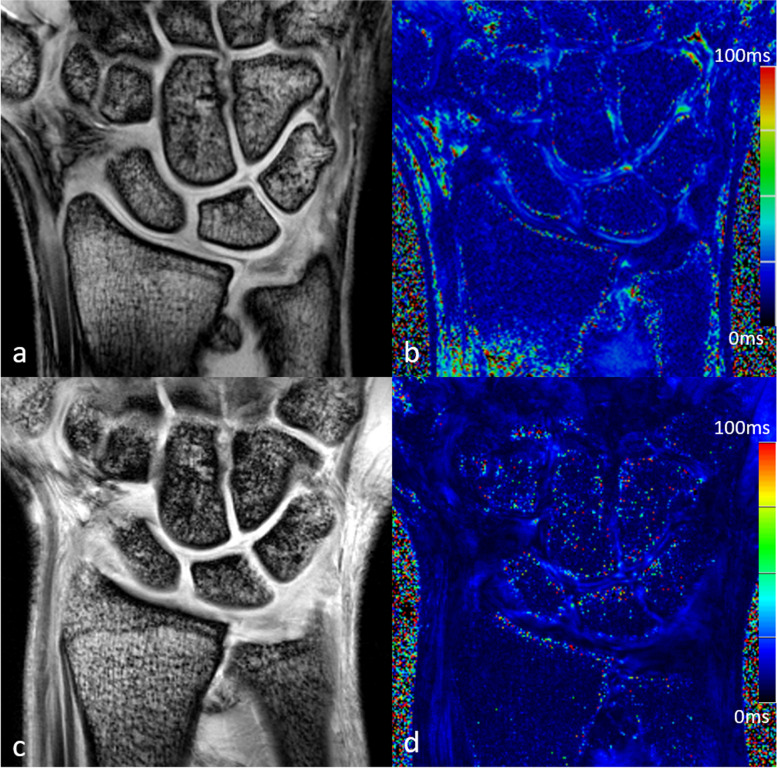
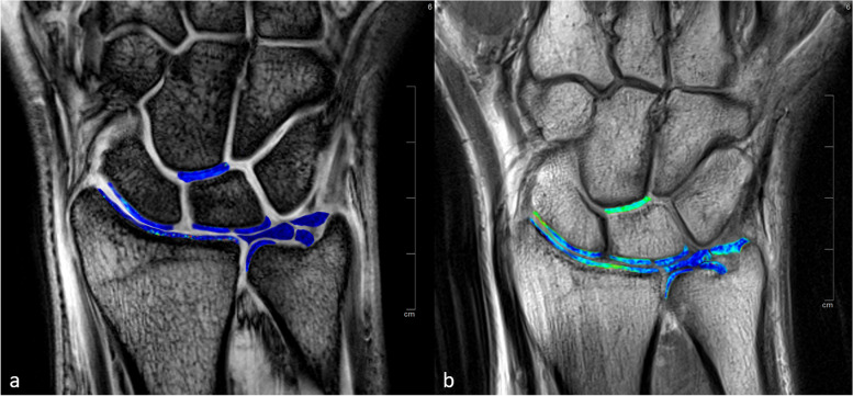
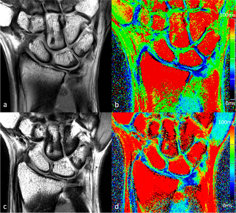
Twenty-five healthy controls and 25 patients with chronic wrist pain were examined at 3 and 7 T on the same day using T2- and T2*-weighted sequences. Six different regions of interest (ROIs) were evaluated for cartilage and 3 ROIs were evaluated at the TFCC based on manual segmentation. Paired t-tests were used to compare T2 and T2* values between field strengths and between different ROIs. Spearman's rank correlation was calculated to assess correlations between T2 and T2* time values at 3 and 7 T.
T2 and T2* time values of the cartilage differed significantly between 3 and 7 T for all ROIs (p ≤ 0.045), with one exception: at the distal lunate, no significant differences in T2 values were observed between field strengths. T2* values differed significantly between 3 and 7 T for all ROIs of the TFCC (p ≤ 0.001). Spearman's rank correlation between 3 and 7 T ranged from 0.03 to 0.62 for T2 values and from 0.01 to 0.48 for T2* values. T2 and T2* values for cartilage varied across anatomic locations in healthy controls at both 3 and 7 T.
Quantitative results of T2 and T2* mapping at the wrist differ between field strengths, with poor correlation between 3 and 7 T. Local variations in cartilage T2 and T2* values are observed in healthy individuals.
T2 and T2* mapping are feasible for compositional imaging of the TFCC and the cartilage at the wrist at both 3 and 7 T, but the clinical interpretation remains challenging due to differences between field strengths and variations between anatomic locations.
•Field strength and anatomic locations influence T2 and T2* values at the wrist. •T2 and T2* values have a poor correlation between 3 and 7 T. •Local reference values are needed for each anatomic location for reliable interpretation.
分析腕关节软骨和三角纤维软骨复合体(TFCC)在 3T 和 7T 时 T2 和 T2*弛豫时间的区域变化,并比较两种场强下的数值。
25 名健康对照者和 25 名慢性腕痛患者于同日在 3T 和 7T 上使用 T2 和 T2加权序列进行检查。基于手动分割,对软骨的 6 个不同感兴趣区(ROI)和 TFCC 的 3 个 ROI 进行评估。使用配对 t 检验比较场强之间和不同 ROI 之间的 T2 和 T2值。使用斯皮尔曼秩相关评估 3T 和 7T 时 T2 和 T2*时间值之间的相关性。
对于所有 ROI(p≤0.045),除了在远端月骨处,3T 和 7T 时软骨的 T2 和 T2时间值均有显著差异,3T 和 7T 时 TFCC 的所有 ROI 的 T2值均有显著差异(p≤0.001)。3T 和 7T 时 T2 值的斯皮尔曼秩相关系数范围为 0.03 至 0.62,T2值的范围为 0.01 至 0.48。在 3T 和 7T 时,健康对照组的软骨 T2 和 T2值在解剖部位上存在差异。
腕部 T2 和 T2图谱的定量结果在两种场强之间存在差异,3T 和 7T 之间相关性较差。在健康个体中观察到软骨 T2 和 T2值的局部变化。
3T 和 7T 时均可行腕部 TFCC 和软骨的 T2 和 T2*成像,但由于场强之间的差异和解剖部位之间的变化,临床解释仍具有挑战性。
•腕部 T2 和 T2值受场强和解剖部位影响。•3T 和 7T 之间 T2 和 T2值相关性较差。•需要为每个解剖部位提供局部参考值,以进行可靠的解释。