Blašković Marko, Butorac Prpić Ivana, Aslan Serhat, Gabrić Dragana, Blašković Dorotea, Cvijanović Peloza Olga, Čandrlić Marija, Perić Kačarević Željka
Dental Clinic Dr. Blašković, Linićeva ulica 16, 51000 Rijeka, Croatia.
Department of Oral Surgery, Faculty of Dental Medicine Rijeka, University of Rijeka, Krešmirova ulica 40/42, 51000 Rijeka, Croatia.
Biomedicines. 2024 Nov 6;12(11):2537. doi: 10.3390/biomedicines12112537.
BACKGROUND/OBJECTIVES: Despite the increased use of new resorbable magnesium membranes, there are no reported cases or studies on the use of resorbable magnesium membranes in combination with bone grafts for alveolar ridge preservation (ARP) in cases with severe buccal bone wall dehiscence. This case report aimed to evaluate the effectiveness of the magnesium membrane shield technique in conjunction with bone grafting for ARP, assessing both clinical outcomes and histological bone regeneration.
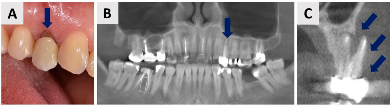
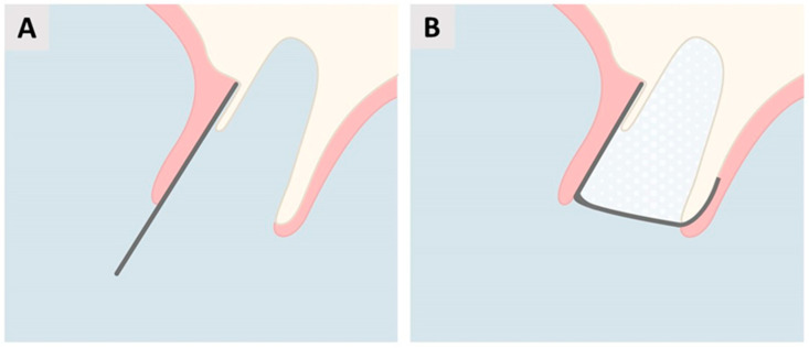
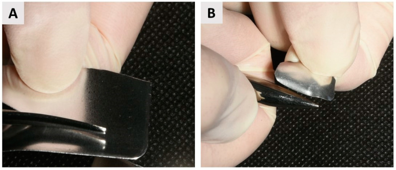
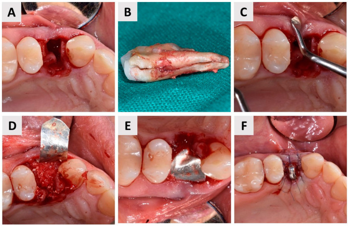
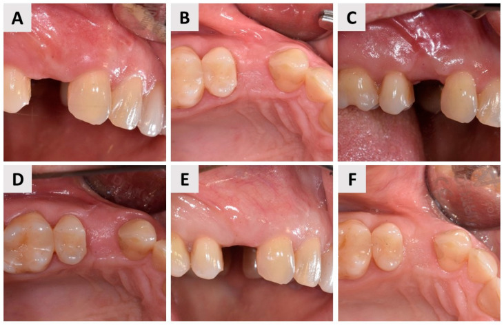
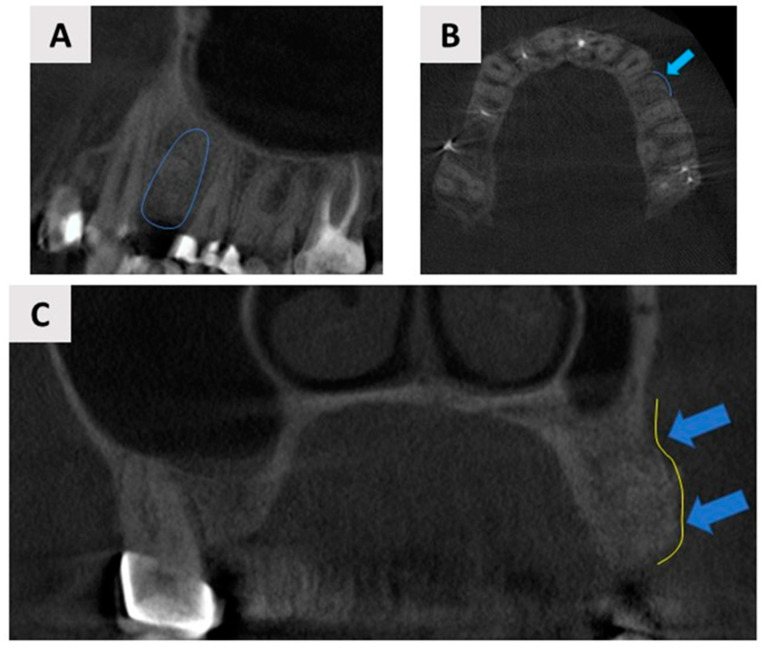
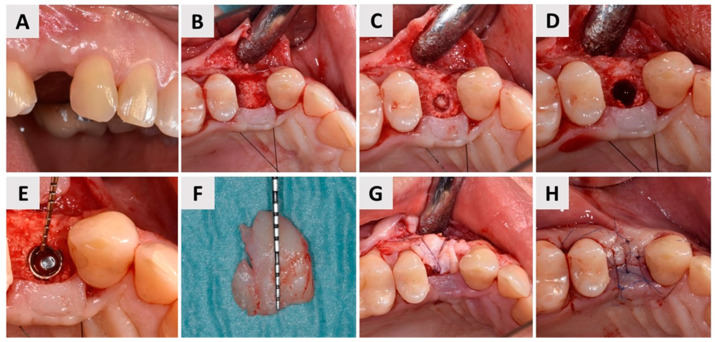
A 44-year-old female patient presented with a vertical fracture on tooth 24 (FDI Notation System) accompanied with complete destruction of the buccal bone wall. The treatment plan included tooth extraction, ARP using a combination of anorganic bovine bone and autologous bone grafting, and the application of a magnesium membrane as a shield to the pre-existing buccal wall. Six months post-procedure, a bone biopsy was taken from the implant site using a trephine bur.
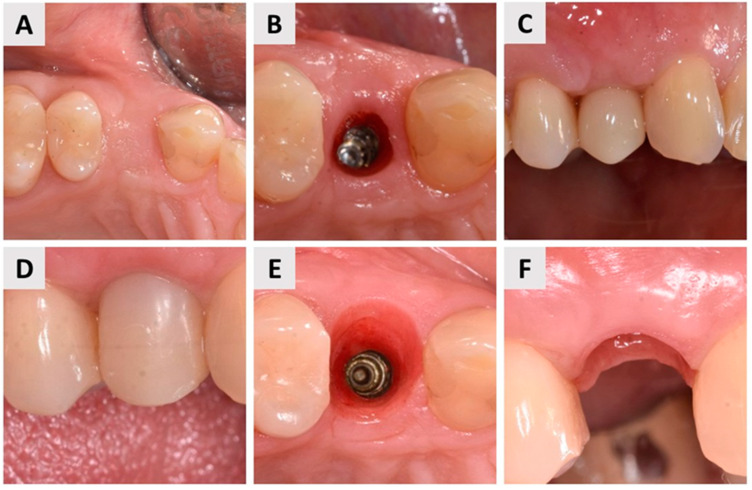
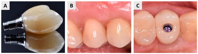
Clinical and radiological evaluations six months after the procedure demonstrated sufficient bone volume for implant placement. Additionally, in the next three months, soft tissue conditioning with a provisional crown resulted in an aesthetically and functionally satisfactory outcome. Histological analysis of the bone biopsy revealed well-formed new bone in direct contact with residual biomaterial, with no signs of inflammation. Osteocytes were clearly visible within the newly formed bone matrix, indicating successful bone maturation. Active osteoblasts were observed along the bone-biomaterial interface, suggesting ongoing bone remodeling and integration. Additionally, histomorphometric evaluation revealed 47% newly formed bone, 32% soft tissue, and 19% residual biomaterial.
This case demonstrates the potential of the magnesium shield technique as an ARP technique in cases with severe buccal wall dehiscence. The technique yielded satisfactory clinical outcomes and promoted successful bone regeneration, as confirmed by histological analysis.
背景/目的:尽管新型可吸收镁膜的使用有所增加,但尚无关于在严重颊侧骨壁裂开病例中使用可吸收镁膜联合骨移植进行牙槽嵴保存(ARP)的病例报告或研究。本病例报告旨在评估镁膜屏障技术联合骨移植用于ARP的有效性,评估临床结果和组织学骨再生情况。
一名44岁女性患者,24号牙(FDI牙位记录系统)发生垂直骨折,伴有颊侧骨壁完全破坏。治疗方案包括拔牙、使用无机牛骨和自体骨移植联合进行ARP,以及在原有颊侧骨壁上应用镁膜作为屏障。术后6个月,使用环钻从种植部位取骨活检样本。
术后6个月的临床和影像学评估显示有足够的骨量用于种植体植入。此外,在接下来的3个月中,使用临时冠进行软组织调整,获得了美观和功能上令人满意的结果。骨活检的组织学分析显示,新形成的骨组织形态良好,与残留生物材料直接接触,无炎症迹象。在新形成的骨基质中可见明显的骨细胞,表明骨成熟成功。在骨-生物材料界面观察到活跃的成骨细胞,提示正在进行骨重塑和整合。此外,组织形态计量学评估显示,新形成骨占47%,软组织占32%,残留生物材料占19%。
本病例证明了镁膜屏障技术作为严重颊侧骨壁裂开病例中ARP技术的潜力。该技术产生了令人满意的临床结果,并促进了成功的骨再生,组织学分析证实了这一点。