Department of Biomedical Sciences, College of Veterinary Medicine and Life Sciences, City University of Hong Kong, 83 Tat Chee Avenue, Kowloon, Hong Kong, China.
Key Laboratory of Biochip Technology, Biotech and Health Centre, Shenzhen Research Institute of City University of Hong Kong, Shenzhen, 518057, China.
Stem Cell Res Ther. 2023 Dec 20;14(1):333. doi: 10.1186/s13287-023-03558-3.
Recent studies demonstrated that elevated osmolarity could induce adipocyte dedifferentiation, representing an appealing procedure to generate multipotent stem cells. Here we aim to elucidate the molecular mechanisms that underlie osmotic induction of adipocyte reprogramming.
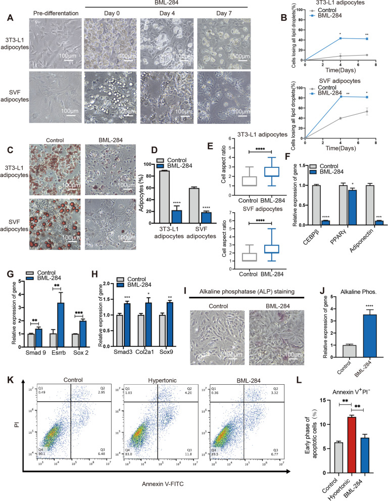
To induce dedifferentiation, the 3T3-L1 or SVF adipocytes were cultured under the hypertonic pressure in 2% PEG 300 medium. Adipocyte dedifferentiation was monitored by aspect ratio measurement, Oil Red staining and qPCR to examine the morphology, lipid droplets, and specific genes of adipocytes, respectively. The osteogenic and chondrogenic re-differentiation capacities of dedifferentiated adipocytes were also examined. To investigate the mechanisms of the osmotic stress-induced dedifferentiation, extracellular vesicles (EVs) were collected from the reprograming cells, followed by proteomic and functional analyses. In addition, qPCR, ELISA, and TNF-α neutralizing antibody (20 ng/ml) was applied to examine the activation and effects of the TNF-α signaling. Furthermore, we also analyzed the Wnt signaling by assessing the activation of β-catenin and applying BML-284, an agonist of β-catenin.
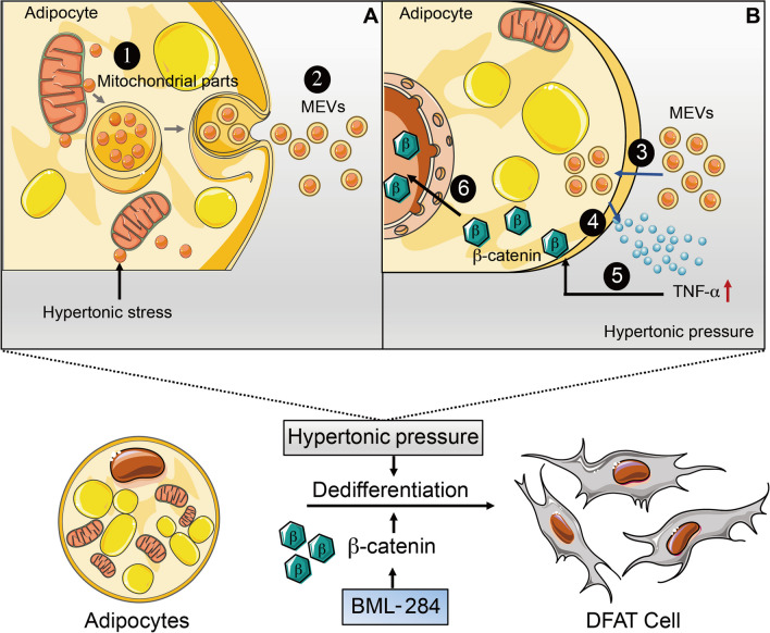
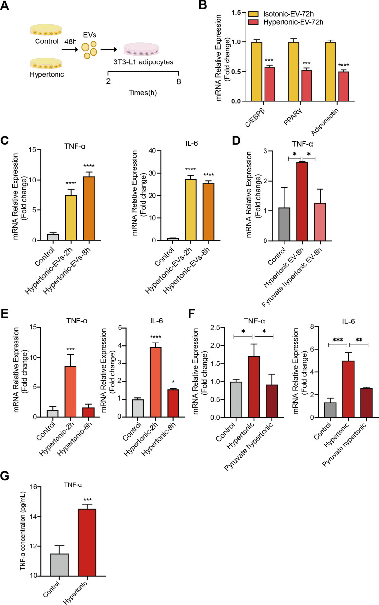
Hypertonic treatment induced dedifferentiation of both 3T3-L1 and the primary stromal vascular fraction (SVF) adipocytes, characterized by morphological and functional changes. Proteomic profiling revealed that hypertonicity induced extracellular vesicles (EVs) containing mitochondrial molecules including NDUFA9 and VDAC. Functionally, the mitochondrial EVs (MEVs) stimulated TNF-α signaling that activates Wnt-β-catenin signaling and adipocyte dedifferentiation. Neutralizing TNF-α inhibited hypertonic dedifferentiation of adipocytes. In addition, direct activation of Wnt-β-catenin signaling using BML-284 could efficiently induce adipocyte dedifferentiation while circumventing the apoptotic effect of the hypertonic treatment.
Hypertonicity prompts the adipocytes to release MEVs, which in turn enhances the secretion of TNF-α as a pro-inflammatory cytokine during the stress response. Importantly, TNF-α is essential for the activation of the Wnt/β-catenin signaling that drives adipocyte dedifferentiation. A caveat of the hypertonic treatment is apoptosis, which could be circumvented by direct activation of the Wnt/β-catenin signaling using BML-284.
最近的研究表明,渗透压升高可诱导脂肪细胞去分化,这代表了一种生成多能干细胞的有吸引力的方法。在这里,我们旨在阐明渗透压诱导脂肪细胞重编程的分子机制。
为了诱导去分化,3T3-L1 或 SVF 脂肪细胞在 2%PEG300 培养基中的高渗压力下培养。通过形态测量、油红染色和 qPCR 分别检测脂肪细胞的形态、脂滴和特定基因,以监测脂肪细胞的去分化。还检查了去分化脂肪细胞的成骨和成软骨再分化能力。为了研究渗透压应激诱导去分化的机制,从重编程细胞中收集细胞外囊泡 (EVs),然后进行蛋白质组学和功能分析。此外,应用 qPCR、ELISA 和 TNF-α 中和抗体(20ng/ml)检测 TNF-α 信号的激活和作用。此外,我们还通过评估β-连环蛋白的激活并应用β-连环蛋白激动剂 BML-284 来分析 Wnt 信号。
高渗处理诱导了 3T3-L1 和原代基质血管部分 (SVF) 脂肪细胞的去分化,其特征在于形态和功能的变化。蛋白质组学分析显示,高渗诱导包含线粒体分子的细胞外囊泡 (EVs),包括 NDUFA9 和 VDAC。功能上,线粒体 EVs (MEVs) 刺激 TNF-α 信号,激活 Wnt-β-连环蛋白信号并诱导脂肪细胞去分化。中和 TNF-α 抑制了脂肪细胞的高渗去分化。此外,使用 BML-284 直接激活 Wnt-β-连环蛋白信号可以有效地诱导脂肪细胞去分化,同时避免高渗处理的凋亡作用。
高渗促使脂肪细胞释放 MEVs,这反过来又增强了应激反应中 TNF-α 作为促炎细胞因子的分泌。重要的是,TNF-α 是激活 Wnt/β-连环蛋白信号的关键,该信号驱动脂肪细胞去分化。高渗处理的一个问题是细胞凋亡,这可以通过使用 BML-284 直接激活 Wnt/β-连环蛋白信号来避免。