Arzanforoosh Fatemeh, Van der Velden Maaike, Berman Avery J L, Van der Voort Sebastian R, Bos Eelke M, Schouten Joost W, Vincent Arnaud J P E, Kros Johan M, Smits Marion, Warnert Esther A H
Department of Radiology & Nuclear Medicine, Erasmus MC, 3015 GD Rotterdam, The Netherlands.
Brain Tumour Center, Erasmus MC Cancer Institute, 3015 GD Rotterdam, The Netherlands.
Cancers (Basel). 2023 Dec 27;16(1):138. doi: 10.3390/cancers16010138.
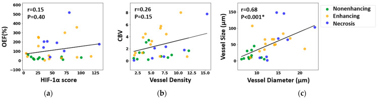
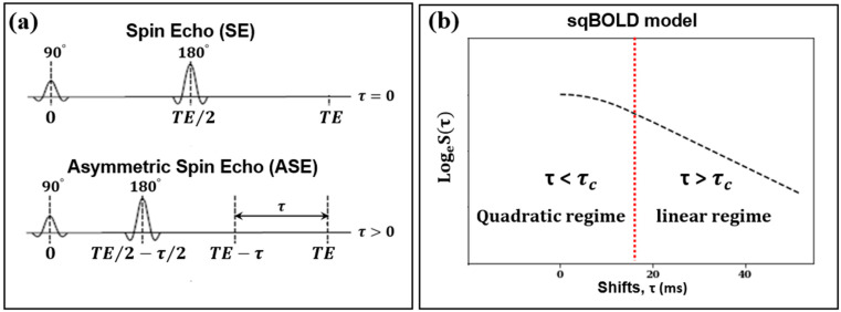
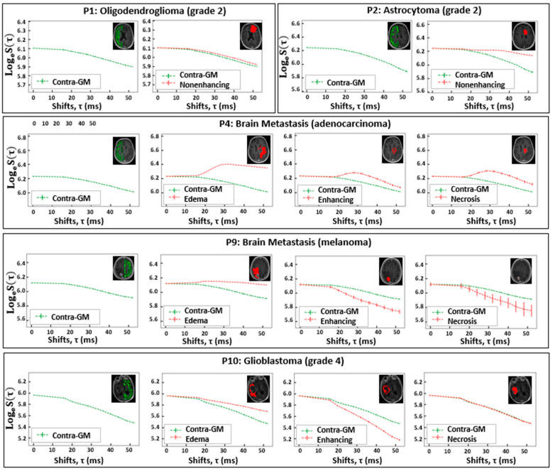
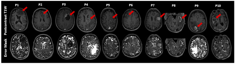
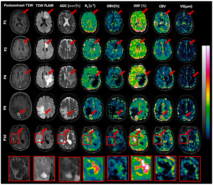
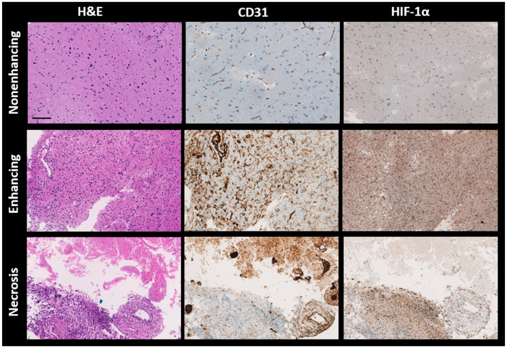
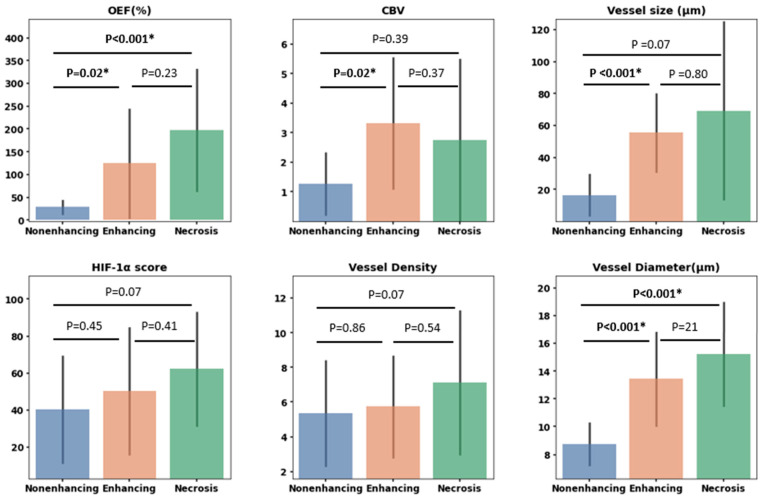
Cerebral hypoxia significantly impacts the progression of brain tumors and their resistance to radiotherapy. This study employed streamlined quantitative blood-oxygen-level-dependent (sqBOLD) MRI to assess the oxygen extraction fraction (OEF)-a measure of how much oxygen is being extracted from vessels, with higher OEF values indicating hypoxia. Simultaneously, we utilized vessel size imaging (VSI) to evaluate microvascular dimensions and blood volume. A cohort of ten patients, divided between those with glioma and those with brain metastases, underwent a 3 Tesla MRI scan. We generated OEF, cerebral blood volume (CBV), and vessel size maps, which guided 3-4 targeted biopsies per patient. Subsequent histological analyses of these biopsies used hypoxia-inducible factor 1-alpha (HIF-1α) for hypoxia and CD31 for microvasculature assessment, followed by a correlation analysis between MRI and histological data. The results showed that while the sqBOLD model was generally applicable to brain tumors, it demonstrated discrepancies in some metastatic tumors, highlighting the need for model adjustments in these cases. The OEF, CBV, and vessel size maps provided insights into the tumor's hypoxic condition, showing intertumoral and intratumoral heterogeneity. A significant relationship between MRI-derived measurements and histological data was only evident in the vessel size measurements (r = 0.68, < 0.001).
脑缺氧显著影响脑肿瘤的进展及其对放疗的抗性。本研究采用简化的定量血氧水平依赖(sqBOLD)磁共振成像来评估氧摄取分数(OEF)——一种衡量从血管中提取多少氧气的指标,OEF值越高表明缺氧。同时,我们利用血管大小成像(VSI)来评估微血管尺寸和血容量。一组十名患者,分为患有胶质瘤的患者和患有脑转移瘤的患者,接受了3特斯拉磁共振成像扫描。我们生成了OEF、脑血容量(CBV)和血管大小图,这些图指导了每位患者进行3 - 4次靶向活检。随后对这些活检组织进行组织学分析,使用缺氧诱导因子1α(HIF - 1α)评估缺氧情况,使用CD31评估微血管情况,然后对磁共振成像和组织学数据进行相关性分析。结果表明,虽然sqBOLD模型通常适用于脑肿瘤,但在一些转移瘤中显示出差异,突出了在这些情况下进行模型调整的必要性。OEF、CBV和血管大小图提供了对肿瘤缺氧状况的见解,显示出肿瘤间和肿瘤内的异质性。仅在血管大小测量中,磁共振成像衍生测量值与组织学数据之间存在显著相关性(r = 0.68,<0.001)。