Van Doorselaer Leen, Verboven Pieter, Nicolai Bart
Mechatronics, Biostatistics and Sensors (MeBioS), Biosystems Department, KU Leuven, Leuven, Belgium.
Flanders Centre of Postharvest Biology, Leuven, Belgium.
Plant Methods. 2024 Jan 19;20(1):12. doi: 10.1186/s13007-024-01137-y.
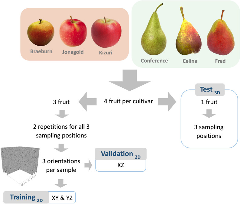
High quality 3D information of the microscopic plant tissue morphology-the spatial organization of cells and intercellular spaces in tissues-helps in understanding physiological processes in a wide variety of plants and tissues. X-ray micro-CT is a valuable tool that is becoming increasingly available in plant research to obtain 3D microstructural information of the intercellular pore space and individual pore sizes and shapes of tissues. However, individual cell morphology is difficult to retrieve from micro-CT as cells cannot be segmented properly due to negligible density differences at cell-to-cell interfaces. To address this, deep learning-based models were trained and tested to segment individual cells using X-ray micro-CT images of parenchyma tissue samples from apple and pear fruit with different cell and porosity characteristics.
The best segmentation model achieved an Aggregated Jaccard Index (AJI) of 0.86 and 0.73 for apple and pear tissue, respectively, which is an improvement over the current benchmark method that achieved AJIs of 0.73 and 0.67. Furthermore, the neural network was able to detect other plant tissue structures such as vascular bundles and stone cell clusters (brachysclereids), of which the latter were shown to strongly influence the spatial organization of pear cells. Based on the AJIs, apple tissue was found to be easier to segment, as the porosity and specific surface area of the pore space are higher and lower, respectively, compared to pear tissue. Moreover, samples with lower pore network connectivity, proved very difficult to segment.
The proposed method can be used to automatically quantify 3D cell morphology of plant tissue from micro-CT instead of opting for laborious manual annotations or less accurate segmentation approaches. In case fruit tissue porosity or pore network connectivity is too low or the specific surface area of the pore space too high, native X-ray micro-CT is unable to provide proper marker points of cell outlines, and one should rely on more elaborate contrast-enhancing scan protocols.
微观植物组织形态的高质量三维信息——组织中细胞和细胞间隙的空间组织——有助于理解多种植物和组织中的生理过程。X射线显微计算机断层扫描(X射线微CT)是一种有价值的工具,在植物研究中越来越多地用于获取细胞间隙孔隙空间以及组织中单个孔隙大小和形状的三维微观结构信息。然而,由于细胞间界面处密度差异可忽略不计,难以从微CT中获取单个细胞形态,因为细胞无法正确分割。为解决这一问题,训练并测试了基于深度学习的模型,以使用具有不同细胞和孔隙率特征的苹果和梨果实薄壁组织样本的X射线微CT图像对单个细胞进行分割。
最佳分割模型对苹果和梨组织的聚合杰卡德指数(AJI)分别达到0.86和0.73,相比当前基准方法(AJI分别为0.73和0.67)有所提高。此外,神经网络能够检测其他植物组织结构,如维管束和石细胞团(短石细胞),其中后者对梨细胞的空间组织有强烈影响。基于AJI,发现苹果组织更容易分割,因为与梨组织相比,孔隙空间的孔隙率和比表面积分别更高和更低。此外,孔隙网络连通性较低的样本分割非常困难。
所提出的方法可用于从微CT自动量化植物组织的三维细胞形态,而无需进行费力的手动注释或不太准确的分割方法。如果果实组织孔隙率或孔隙网络连通性过低,或者孔隙空间的比表面积过高,原生X射线微CT无法提供细胞轮廓的适当标记点,则应依赖更精细的对比度增强扫描方案。