• 文献检索
  • 文档翻译
  • 深度研究
  • 学术资讯
  • Suppr Zotero 插件Zotero 插件
  • 邀请有礼
  • 套餐&价格
  • 历史记录
应用&插件
Suppr Zotero 插件Zotero 插件浏览器插件Mac 客户端Windows 客户端微信小程序
定价
高级版会员购买积分包购买API积分包
服务
文献检索文档翻译深度研究API 文档MCP 服务
关于我们
关于 Suppr公司介绍联系我们用户协议隐私条款
关注我们

Suppr 超能文献

核心技术专利:CN118964589B侵权必究
粤ICP备2023148730 号-1Suppr @ 2026

文献检索

告别复杂PubMed语法,用中文像聊天一样搜索,搜遍4000万医学文献。AI智能推荐,让科研检索更轻松。

立即免费搜索

文件翻译

保留排版,准确专业,支持PDF/Word/PPT等文件格式,支持 12+语言互译。

免费翻译文档

深度研究

AI帮你快速写综述,25分钟生成高质量综述,智能提取关键信息,辅助科研写作。

立即免费体验

以细胞分辨率准确且灵活地对植物组织进行三维分割。

Accurate and versatile 3D segmentation of plant tissues at cellular resolution.

机构信息

Heidelberg Collaboratory for Image Processing, Heidelberg University, Heidelberg, Germany.

EMBL, Heidelberg, Germany.

出版信息

Elife. 2020 Jul 29;9:e57613. doi: 10.7554/eLife.57613.

DOI:10.7554/eLife.57613
PMID:32723478
原文链接:https://pmc.ncbi.nlm.nih.gov/articles/PMC7447435/
Abstract

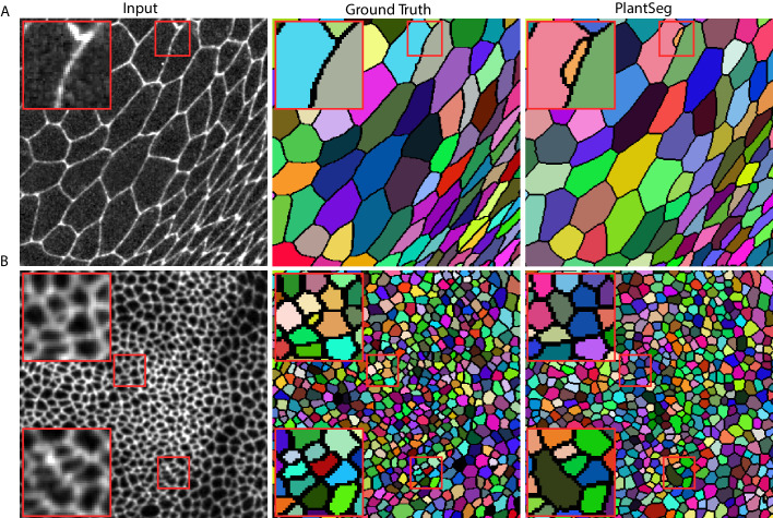
Quantitative analysis of plant and animal morphogenesis requires accurate segmentation of individual cells in volumetric images of growing organs. In the last years, deep learning has provided robust automated algorithms that approach human performance, with applications to bio-image analysis now starting to emerge. Here, we present PlantSeg, a pipeline for volumetric segmentation of plant tissues into cells. PlantSeg employs a convolutional neural network to predict cell boundaries and graph partitioning to segment cells based on the neural network predictions. PlantSeg was trained on fixed and live plant organs imaged with confocal and light sheet microscopes. PlantSeg delivers accurate results and generalizes well across different tissues, scales, acquisition settings even on non plant samples. We present results of PlantSeg applications in diverse developmental contexts. PlantSeg is free and open-source, with both a command line and a user-friendly graphical interface.

摘要

植物和动物形态发生的定量分析需要对生长器官的体积图像中的单个细胞进行精确分割。在过去的几年中,深度学习提供了强大的自动化算法,可以达到人类的表现水平,现在生物图像分析的应用也开始出现。在这里,我们提出了 PlantSeg,这是一个将植物组织分割成细胞的体积分割管道。PlantSeg 使用卷积神经网络来预测细胞边界,并使用图划分根据神经网络预测来分割细胞。PlantSeg 是在使用共聚焦和光片显微镜成像的固定和活体植物器官上进行训练的。PlantSeg 提供了准确的结果,并可以很好地推广到不同的组织、尺度、采集设置,甚至是非植物样本。我们展示了 PlantSeg 在不同发育背景下的应用结果。PlantSeg 是免费的开源软件,同时提供命令行和用户友好的图形界面。

https://cdn.ncbi.nlm.nih.gov/pmc/blobs/871a/7447435/e9d79129ad6d/elife-57613-app6-fig1.jpg
https://cdn.ncbi.nlm.nih.gov/pmc/blobs/871a/7447435/f6d3397c8e57/elife-57613-fig1.jpg
https://cdn.ncbi.nlm.nih.gov/pmc/blobs/871a/7447435/3c938615822e/elife-57613-fig2.jpg
https://cdn.ncbi.nlm.nih.gov/pmc/blobs/871a/7447435/7f48432612bb/elife-57613-fig3.jpg
https://cdn.ncbi.nlm.nih.gov/pmc/blobs/871a/7447435/069733e9cb40/elife-57613-fig4.jpg
https://cdn.ncbi.nlm.nih.gov/pmc/blobs/871a/7447435/15e3d74f8525/elife-57613-fig5.jpg
https://cdn.ncbi.nlm.nih.gov/pmc/blobs/871a/7447435/09ee7468199d/elife-57613-fig6.jpg
https://cdn.ncbi.nlm.nih.gov/pmc/blobs/871a/7447435/ca3ce8644c9a/elife-57613-fig7.jpg
https://cdn.ncbi.nlm.nih.gov/pmc/blobs/871a/7447435/9f75f5b8249f/elife-57613-fig8.jpg
https://cdn.ncbi.nlm.nih.gov/pmc/blobs/871a/7447435/950153d2b8d0/elife-57613-fig9.jpg
https://cdn.ncbi.nlm.nih.gov/pmc/blobs/871a/7447435/53a981b1dcb3/elife-57613-fig10.jpg
https://cdn.ncbi.nlm.nih.gov/pmc/blobs/871a/7447435/877f3b4370ae/elife-57613-app1-fig1.jpg
https://cdn.ncbi.nlm.nih.gov/pmc/blobs/871a/7447435/473eec5f84cd/elife-57613-app3-fig1.jpg
https://cdn.ncbi.nlm.nih.gov/pmc/blobs/871a/7447435/8c2fce17b2ae/elife-57613-app4-fig1.jpg
https://cdn.ncbi.nlm.nih.gov/pmc/blobs/871a/7447435/e9d79129ad6d/elife-57613-app6-fig1.jpg

相似文献

1
Accurate and versatile 3D segmentation of plant tissues at cellular resolution.以细胞分辨率准确且灵活地对植物组织进行三维分割。
Elife. 2020 Jul 29;9:e57613. doi: 10.7554/eLife.57613.
2
A deep learning-based toolkit for 3D nuclei segmentation and quantitative analysis in cellular and tissue context.基于深度学习的 3D 细胞核分割和细胞及组织定量分析工具包。
Development. 2024 Jul 15;151(14). doi: 10.1242/dev.202800. Epub 2024 Jul 18.
3
A robust transformer-based pipeline of 3D cell alignment, denoise and instance segmentation on electron microscopy sequence images.基于稳健的变压器的 3D 细胞配准、去噪和电子显微镜序列图像实例分割流水线。
J Plant Physiol. 2024 Jun;297:154236. doi: 10.1016/j.jplph.2024.154236. Epub 2024 Apr 2.
4
CellSeg: a robust, pre-trained nucleus segmentation and pixel quantification software for highly multiplexed fluorescence images.CellSeg:一款用于高度多重荧光图像的强大的、预训练的细胞核分割和像素定量软件。
BMC Bioinformatics. 2022 Jan 18;23(1):46. doi: 10.1186/s12859-022-04570-9.
5
Label-free cell segmentation of diverse lymphoid tissues in 2D and 3D.二维和三维空间中多样化淋巴组织的无标记细胞分割。
Cell Rep Methods. 2023 Feb 2;3(2):100398. doi: 10.1016/j.crmeth.2023.100398. eCollection 2023 Feb 27.
6
DeepImageTranslator: A free, user-friendly graphical interface for image translation using deep-learning and its applications in 3D CT image analysis.深度图像翻译器:一个免费、用户友好的图形界面,用于利用深度学习进行图像翻译及其在三维CT图像分析中的应用。
SLAS Technol. 2022 Feb;27(1):76-84. doi: 10.1016/j.slast.2021.10.014. Epub 2021 Oct 25.
7
Deep convolutional neural network for segmentation of thoracic organs-at-risk using cropped 3D images.使用裁剪的 3D 图像进行胸部危险器官分割的深度卷积神经网络。
Med Phys. 2019 May;46(5):2169-2180. doi: 10.1002/mp.13466. Epub 2019 Mar 21.
8
3D APA-Net: 3D Adversarial Pyramid Anisotropic Convolutional Network for Prostate Segmentation in MR Images.3DAPA-Net:基于三维对抗金字塔各向异性卷积网络的磁共振图像前列腺分割。
IEEE Trans Med Imaging. 2020 Feb;39(2):447-457. doi: 10.1109/TMI.2019.2928056. Epub 2019 Jul 11.
9
CellECT: cell evolution capturing tool.CellECT:细胞进化捕获工具。
BMC Bioinformatics. 2016 Feb 17;17:88. doi: 10.1186/s12859-016-0927-7.
10
Single-cell segmentation in bacterial biofilms with an optimized deep learning method enables tracking of cell lineages and measurements of growth rates.利用优化的深度学习方法对细菌生物膜中的单细胞进行分割,可实现细胞谱系追踪和生长速率测量。
Mol Microbiol. 2023 Jun;119(6):659-676. doi: 10.1111/mmi.15064. Epub 2023 Apr 17.

引用本文的文献

1
Single cell resolution 3D imaging and segmentation within intact live tissues.完整活组织内的单细胞分辨率三维成像与分割
Npj Imaging. 2025 Sep 3;3(1):40. doi: 10.1038/s44303-025-00099-7.
2
Ultrack: pushing the limits of cell tracking across biological scales.Ultrack:跨越生物尺度推动细胞追踪的极限。
Nat Methods. 2025 Aug 25. doi: 10.1038/s41592-025-02778-0.
3
MRI-derived quantification of hepatic vessel-to-volume ratios in chronic liver disease using a deep learning approach.使用深度学习方法对慢性肝病中肝脏血管与体积比进行MRI衍生量化

本文引用的文献

1
Generalised Dice Overlap as a Deep Learning Loss Function for Highly Unbalanced Segmentations.广义骰子重叠作为高度不平衡分割的深度学习损失函数
Deep Learn Med Image Anal Multimodal Learn Clin Decis Support (2017). 2017;2017:240-248. doi: 10.1007/978-3-319-67558-9_28. Epub 2017 Sep 9.
2
BigDataProcessor2: a free and open-source Fiji plugin for inspection and processing of TB sized image data.BigDataProcessor2:一款免费的开源 Fiji 插件,用于检查和处理 TB 级图像数据。
Bioinformatics. 2021 Sep 29;37(18):3079-3081. doi: 10.1093/bioinformatics/btab106.
3
Protocol for rapid clearing and staining of fixed Arabidopsis ovules for improved imaging by confocal laser scanning microscopy.
Eur Radiol Exp. 2025 Aug 12;9(1):75. doi: 10.1186/s41747-025-00612-y.
4
The pipelines of deep learning-based plant image processing.基于深度学习的植物图像处理流程。
Quant Plant Biol. 2025 Jul 25;6:e23. doi: 10.1017/qpb.2025.10018. eCollection 2025.
5
Deep Neural Networks to Register and Annotate Cells in Moving and Deforming Nervous Systems.用于在移动和变形神经系统中对细胞进行配准和注释的深度神经网络。
bioRxiv. 2025 Jun 21:2024.07.18.601886. doi: 10.1101/2024.07.18.601886.
6
Plants monitor the integrity of their barrier by sensing gas diffusion.植物通过感知气体扩散来监测其屏障的完整性。
Nature. 2025 Jul 2. doi: 10.1038/s41586-025-09223-4.
7
A Method to Visualize Cell Proliferation of : A Case Study of the Root Apical Meristem.一种可视化细胞增殖的方法:以根尖分生组织为例
Plant Direct. 2025 Apr 28;9(4):e70060. doi: 10.1002/pld3.70060. eCollection 2025 Apr.
8
A high-throughput differential chemical genetic screen uncovers genotype-specific compounds altering plant growth.一项高通量差异化学遗传筛选发现了改变植物生长的基因型特异性化合物。
iScience. 2025 Apr 8;28(5):112375. doi: 10.1016/j.isci.2025.112375. eCollection 2025 May 16.
9
In toto live imaging of Erk signaling dynamics in developing zebrafish hepatocytes.斑马鱼发育中肝细胞内Erk信号动力学的全时程活体成像
Dev Biol. 2025 Jul;523:43-50. doi: 10.1016/j.ydbio.2025.04.009. Epub 2025 Apr 12.
10
Point-cloud segmentation with in-silico data augmentation for prostate cancer treatment.用于前列腺癌治疗的基于计算机模拟数据增强的点云分割
Med Phys. 2025 Apr 3. doi: 10.1002/mp.17815.
用于通过共聚焦激光扫描显微镜改善成像的拟南芥固定胚珠快速清除和染色方案。
Plant Methods. 2019 Oct 25;15:120. doi: 10.1186/s13007-019-0505-x. eCollection 2019.
4
ilastik: interactive machine learning for (bio)image analysis.ilastik:用于(生物)图像处理的交互式机器学习。
Nat Methods. 2019 Dec;16(12):1226-1232. doi: 10.1038/s41592-019-0582-9. Epub 2019 Sep 30.
5
Cytoskeleton Dynamics Are Necessary for Early Events of Lateral Root Initiation in Arabidopsis.细胞骨架动力学对于拟南芥侧根起始的早期事件是必要的。
Curr Biol. 2019 Aug 5;29(15):2443-2454.e5. doi: 10.1016/j.cub.2019.06.039. Epub 2019 Jul 18.
6
Quantifying Plant Growth and Cell Proliferation with MorphoGraphX.使用MorphoGraphX量化植物生长和细胞增殖。
Methods Mol Biol. 2019;1992:269-290. doi: 10.1007/978-1-4939-9469-4_18.
7
Deep learning for cellular image analysis.深度学习在细胞图像分析中的应用。
Nat Methods. 2019 Dec;16(12):1233-1246. doi: 10.1038/s41592-019-0403-1. Epub 2019 May 27.
8
A Growth-Based Framework for Leaf Shape Development and Diversity.基于生长的叶片形状发育和多样性框架。
Cell. 2019 May 30;177(6):1405-1418.e17. doi: 10.1016/j.cell.2019.05.011. Epub 2019 May 23.
9
Learn to segment single cells with deep distance estimator and deep cell detector.学习使用深度距离估计器和深度细胞探测器对单细胞进行分割。
Comput Biol Med. 2019 May;108:133-141. doi: 10.1016/j.compbiomed.2019.04.006. Epub 2019 Apr 8.
10
U-Net: deep learning for cell counting, detection, and morphometry.U-Net:用于细胞计数、检测和形态测量学的深度学习。
Nat Methods. 2019 Jan;16(1):67-70. doi: 10.1038/s41592-018-0261-2. Epub 2018 Dec 17.