Choi Joon Yul, Ryu Ik Hee, Kim Jin Kuk, Lee In Sik, Yoo Tae Keun
Department of Biomedical Engineering, Yonsei University, Wonju, South Korea.
Department of Refractive Surgery, B&VIIT Eye Center, B2 GT Tower, 1317-23 Seocho-Dong, Seocho-Gu, Seoul, South Korea.
BMC Med Inform Decis Mak. 2024 Jan 26;24(1):25. doi: 10.1186/s12911-024-02431-4.
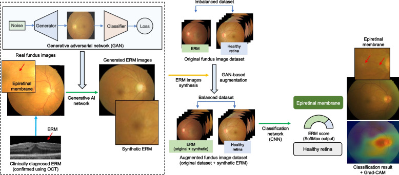
The epiretinal membrane (ERM) is a common retinal disorder characterized by abnormal fibrocellular tissue at the vitreomacular interface. Most patients with ERM are asymptomatic at early stages. Therefore, screening for ERM will become increasingly important. Despite the high prevalence of ERM, few deep learning studies have investigated ERM detection in the color fundus photography (CFP) domain. In this study, we built a generative model to enhance ERM detection performance in the CFP.
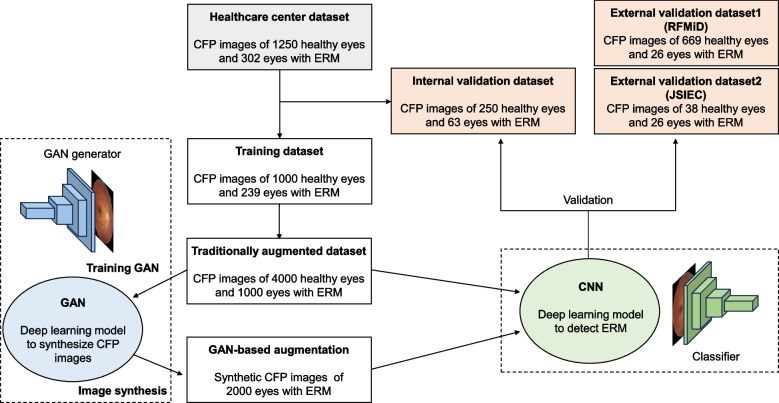
This deep learning study retrospectively collected 302 ERM and 1,250 healthy CFP data points from a healthcare center. The generative model using StyleGAN2 was trained using single-center data. EfficientNetB0 with StyleGAN2-based augmentation was validated using independent internal single-center data and external datasets. We randomly assigned healthcare center data to the development (80%) and internal validation (20%) datasets. Data from two publicly accessible sources were used as external validation datasets.
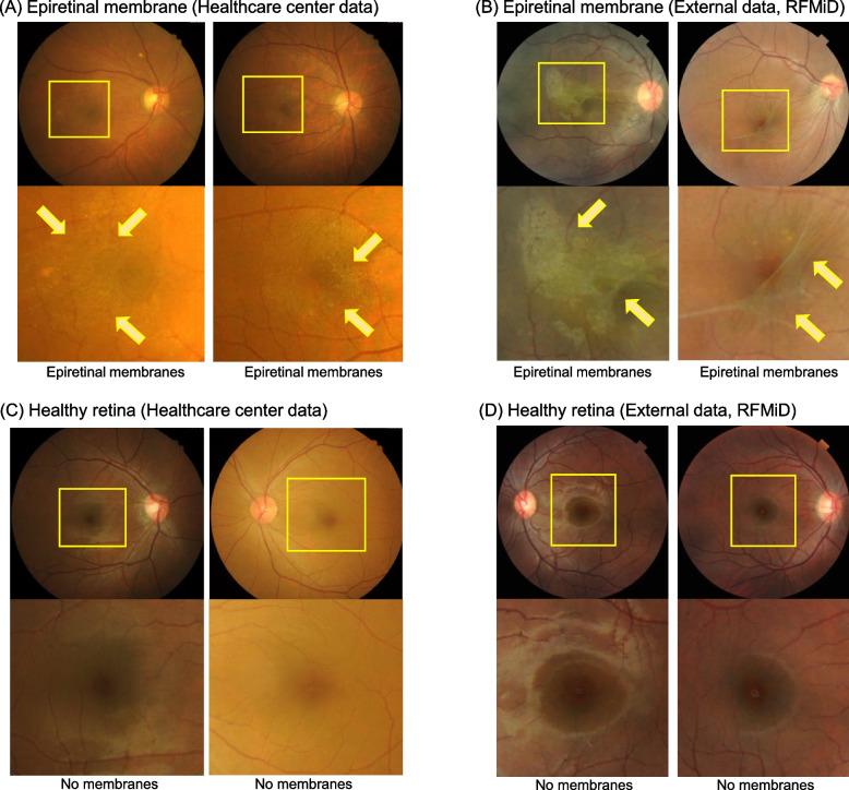
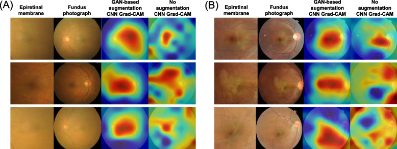
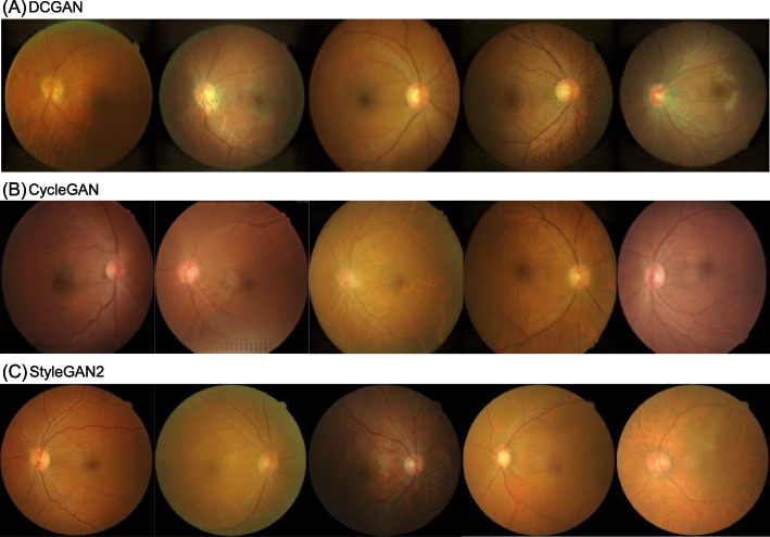
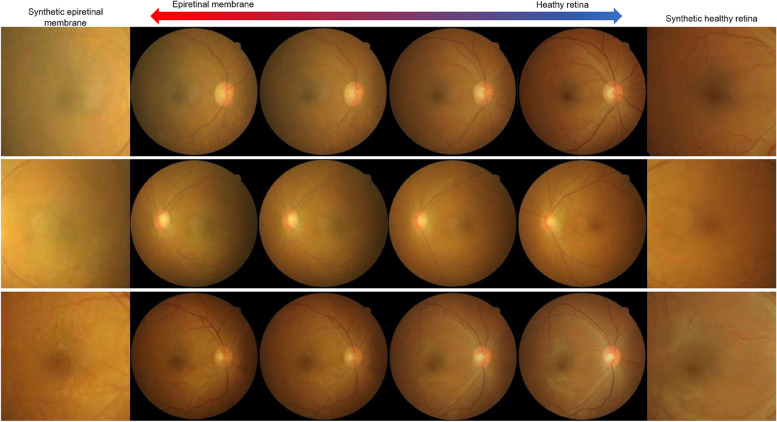
StyleGAN2 facilitated realistic CFP synthesis with the characteristic cellophane reflex features of the ERM. The proposed method with StyleGAN2-based augmentation outperformed the typical transfer learning without a generative adversarial network. The proposed model achieved an area under the receiver operating characteristic (AUC) curve of 0.926 for internal validation. AUCs of 0.951 and 0.914 were obtained for the two external validation datasets. Compared with the deep learning model without augmentation, StyleGAN2-based augmentation improved the detection performance and contributed to the focus on the location of the ERM.
We proposed an ERM detection model by synthesizing realistic CFP images with the pathological features of ERM through generative deep learning. We believe that our deep learning framework will help achieve a more accurate detection of ERM in a limited data setting.
视网膜前膜(ERM)是一种常见的视网膜疾病,其特征是玻璃体黄斑界面出现异常的纤维细胞组织。大多数ERM患者在早期无症状。因此,ERM筛查将变得越来越重要。尽管ERM的患病率很高,但很少有深度学习研究在彩色眼底照片(CFP)领域研究ERM检测。在本研究中,我们构建了一个生成模型以提高CFP中ERM的检测性能。
这项深度学习研究回顾性地从一个医疗中心收集了302个ERM和1250个健康CFP数据点。使用StyleGAN2的生成模型使用单中心数据进行训练。基于StyleGAN2增强的EfficientNetB0使用独立的内部单中心数据和外部数据集进行验证。我们将医疗中心的数据随机分配到开发(80%)和内部验证(20%)数据集。来自两个可公开获取来源的数据用作外部验证数据集。
StyleGAN2促进了具有ERM特征性玻璃纸样反光特征的逼真CFP合成。所提出的基于StyleGAN2增强的方法优于没有生成对抗网络的典型迁移学习。所提出的模型在内部验证中实现了受试者操作特征(AUC)曲线下面积为0.926。两个外部验证数据集的AUC分别为0.951和0.914。与没有增强的深度学习模型相比,基于StyleGAN2的增强提高了检测性能,并有助于关注ERM的位置。
我们通过生成式深度学习合成具有ERM病理特征的逼真CFP图像,提出了一种ERM检测模型。我们相信我们的深度学习框架将有助于在有限的数据设置中更准确地检测ERM。