Wang Haojue, Xiang Dajun, Lu Xianyi, Fang Ling, Cui Chengjun, Shi Qifeng, Yang Xiaojun
Department of Obstetrics and Gynecology, Wuxi Xishan People's Hospital, (Wuxi Branch of Zhongda Hospital Southeast University), 214015, Wuxi, Jiangsu, China.
The First Affiliated Hospital of Soochow University, Suzhou, Jiangsu, 215006, China.
Heliyon. 2024 Jan 11;10(2):e24460. doi: 10.1016/j.heliyon.2024.e24460. eCollection 2024 Jan 30.
Cervical cancer (CC) is currently the most common malignant tumour in the female reproductive tract, and paclitaxel (PTX) is a commonly used chemotherapeutic agent, but tumour cell resistance will seriously affect the therapeutic efficacy of PTX. Nanoparticle human serum albumin-bound paclitaxel (Nano-HSA-PTX) is a novel drug delivery modality that may have superior effects to PTX alone.
To clarify the effect of Nano-HSA-PTX on cervical carcinoma (CC) cells and the underlying mechanisms.
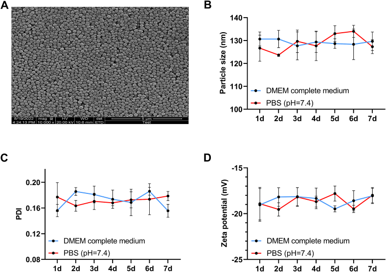
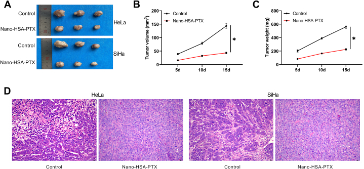
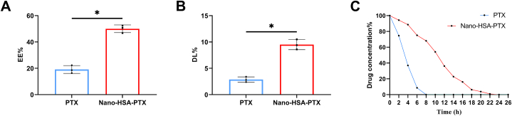
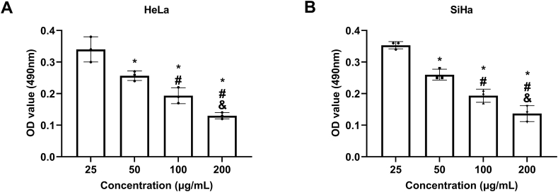
After the preparation of Nano-HSA-PTX, its morphology was observed by electron transmission microscope (TEM), and its entrapment efficiency (EE%) and drug loading rate (DL%) were detected. Nano-HSA-PTX was compared with conventional PTX for drug metabolism. Additionally, CC HeLa and SiHa cells were purchased and divided into three groups to treat with Nano-HSA-PTX, PTX and normal saline, respectively. MTT, cell cloning, Transwell and cell scratch assays were carried out to determine cell proliferation, invasion and migration, flow cytometry and Western blotting were performed to detect apoptosis rate and apoptosis-related protein expression, and PCR was conducted to quantify oxidative damage indicators. Further, CYP3A4 and CYP2C8 expression patterns in CC cells (HeLa and SiHa) and human normal cervical epithelia (End1/E6E7) and the changes of their levels under the intervention of Nano-HSA-PTX were measured. Subsequently, C57BL/6mice were purchased for subcutaneous tumorigenesis experiment to observe the impact of Nano-HSA-PTX on tumor growth.
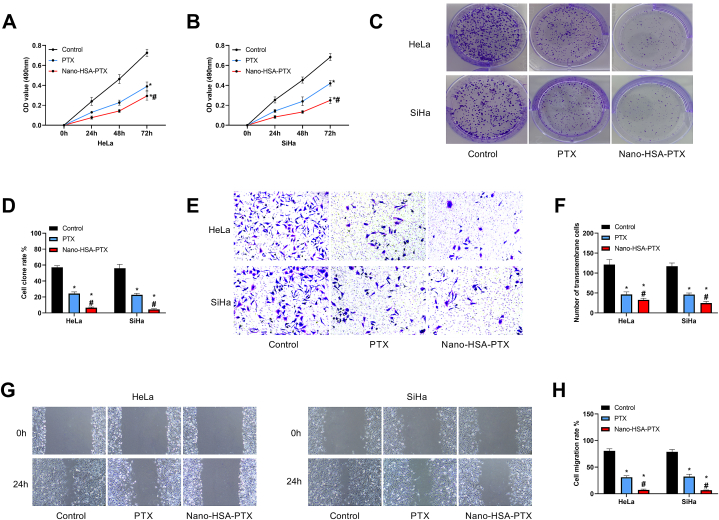
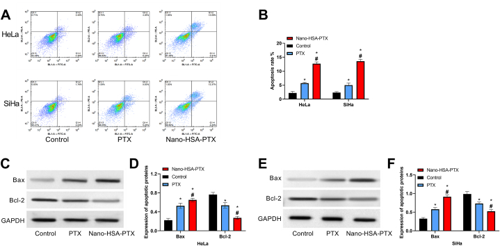
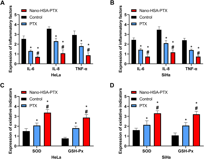
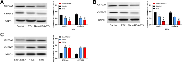
Under TEM, Nano-HSA-PTX was complete and arranged compactly, with a stable structure and markedly higher EE% and DL% than PTX (P < 0.05). Under Nano-HSA-PTX intervention, the proliferation, invasion, migration and oxidative damage of HeLa and SiHa were significantly decreased compared with the control and PTX groups, while the apoptosis was increased (P < 0.05). Besides, elevated CYP3A4 and CYP2C8 levels were observed in CC cells, which were inhibited by Nano-HSA-PTX and PTX (P < 0.05). Finally, tumorigenesis experiments in nude mice revealed that Nano-HSA-PTX could inhibit tumor growth.
Compared with PTX, Nano-HSA-PTX has a superior effect of inhibiting CC activity. And this mechanism of action was carried out by inhibiting the expression of CYP3A4 and CYP2C8.
宫颈癌(CC)是目前女性生殖道最常见的恶性肿瘤,紫杉醇(PTX)是常用的化疗药物,但肿瘤细胞耐药会严重影响PTX的治疗效果。纳米白蛋白结合紫杉醇(Nano-HSA-PTX)是一种新型给药方式,可能比单纯PTX具有更优效果。
阐明Nano-HSA-PTX对宫颈癌细胞的作用及潜在机制。
制备Nano-HSA-PTX后,通过透射电子显微镜(TEM)观察其形态,检测其包封率(EE%)和载药率(DL%)。将Nano-HSA-PTX与传统PTX进行药物代谢比较。此外,购买CC HeLa和SiHa细胞,分为三组,分别用Nano-HSA-PTX、PTX和生理盐水处理。进行MTT、细胞克隆、Transwell和细胞划痕试验以确定细胞增殖、侵袭和迁移,进行流式细胞术和蛋白质免疫印迹法检测凋亡率和凋亡相关蛋白表达,并进行PCR定量氧化损伤指标。进一步检测CC细胞(HeLa和SiHa)和人正常宫颈上皮(End1/E6E7)中CYP3A4和CYP2C8的表达模式以及Nano-HSA-PTX干预下其水平的变化。随后,购买C57BL/6小鼠进行皮下成瘤实验,观察Nano-HSA-PTX对肿瘤生长的影响。
在TEM下,Nano-HSA-PTX完整且排列紧密,结构稳定,EE%和DL%明显高于PTX(P < 0.05)。在Nano-HSA-PTX干预下,与对照组和PTX组相比,HeLa和SiHa的增殖、侵袭、迁移和氧化损伤明显降低,而凋亡增加(P < 0.05)。此外,在CC细胞中观察到CYP3A4和CYP2C8水平升高,Nano-HSA-PTX和PTX可抑制其升高(P < 0.05)。最后,裸鼠成瘤实验表明Nano-HSA-PTX可抑制肿瘤生长。
与PTX相比,Nano-HSA-PTX具有更优的抑制CC活性的效果。且这种作用机制是通过抑制CYP3A4和CYP2C8的表达来实现的。