Mishra Zubin, Wang Ziyuan, Xu Emily, Xu Sophia, Majid Iyad, Sadda SriniVas R, Hu Zhihong Jewel
Doheny Image Analysis Laboratory, Doheny Eye Institute, Pasadena, CA, 91103, USA.
Case Western Reserve University School of Medicine, Cleveland, OH, 44106, USA.
medRxiv. 2024 Feb 13:2024.02.11.24302670. doi: 10.1101/2024.02.11.24302670.
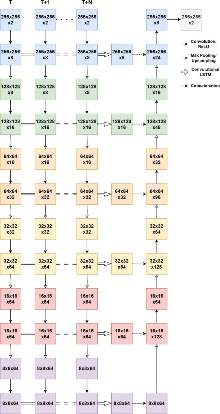
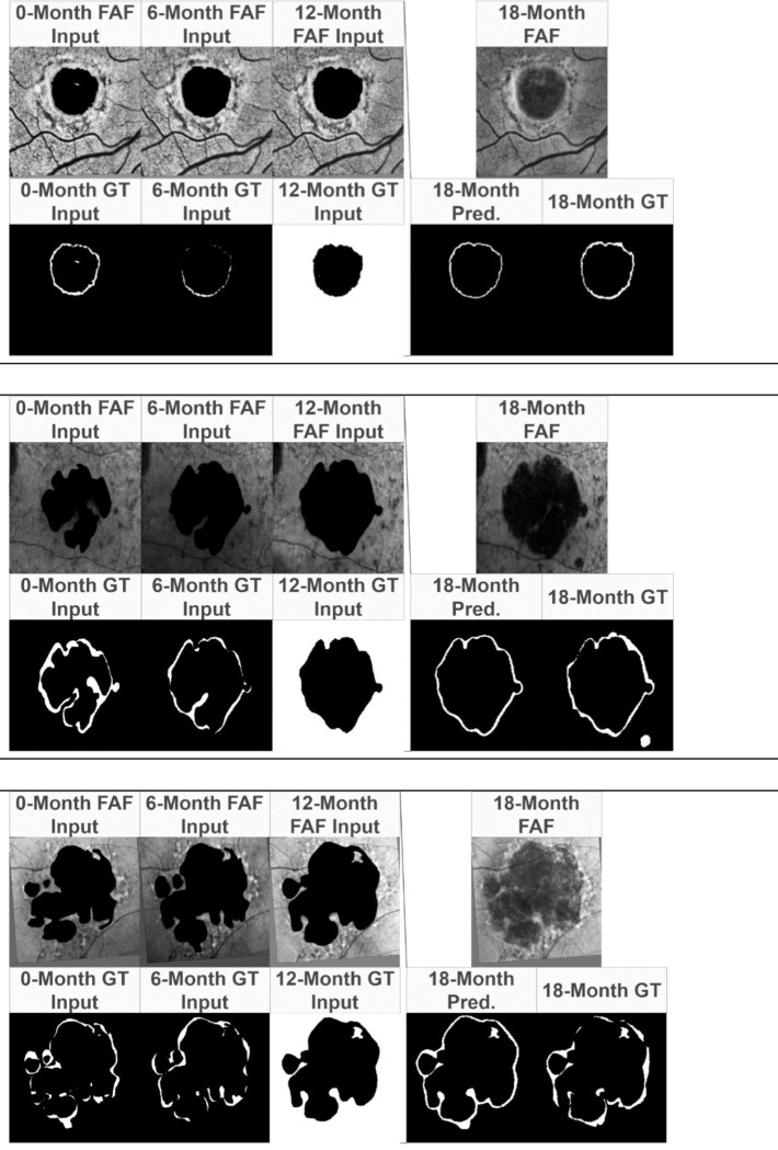
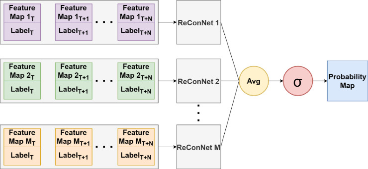
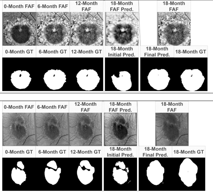
Stargardt disease and age-related macular degeneration are the leading causes of blindness in the juvenile and geriatric populations, respectively. The formation of atrophic regions of the macula is a hallmark of the end-stages of both diseases. The progression of these diseases is tracked using various imaging modalities, two of the most common being fundus autofluorescence (FAF) imaging and spectral-domain optical coherence tomography (SD-OCT). This study seeks to investigate the use of longitudinal FAF and SD-OCT imaging (month 0, month 6, month 12, and month 18) data for the predictive modelling of future atrophy in Stargardt and geographic atrophy. To achieve such an objective, we develop a set of novel deep convolutional neural networks enhanced with recurrent network units for longitudinal prediction and concurrent learning of ensemble network units (termed ReConNet) which take advantage of improved retinal layer features beyond the mean intensity features. Using FAF images, the neural network presented in this paper achieved mean (± standard deviation, SD) and median Dice coefficients of 0.895 (± 0.086) and 0.922 for Stargardt atrophy, and 0.864 (± 0.113) and 0.893 for geographic atrophy. Using SD-OCT images for Stargardt atrophy, the neural network achieved mean and median Dice coefficients of 0.882 (± 0.101) and 0.906, respectively. When predicting only the interval growth of the atrophic lesions with FAF images, mean (± SD) and median Dice coefficients of 0.557 (± 0.094) and 0.559 were achieved for Stargardt atrophy, and 0.612 (± 0.089) and 0.601 for geographic atrophy. The prediction performance in OCT images is comparably good to that using FAF which opens a new, more efficient, and practical door in the assessment of atrophy progression for clinical trials and retina clinics, beyond widely used FAF. These results are highly encouraging for a high-performance interval growth prediction when more frequent or longer-term longitudinal data are available in our clinics. This is a pressing task for our next step in ongoing research.
斯塔加特病和年龄相关性黄斑变性分别是青少年和老年人群失明的主要原因。黄斑萎缩区域的形成是这两种疾病末期的一个标志。使用各种成像方式来追踪这些疾病的进展,其中两种最常见的是眼底自发荧光(FAF)成像和光谱域光学相干断层扫描(SD-OCT)。本研究旨在调查使用纵向FAF和SD-OCT成像(第0个月、第6个月、第12个月和第18个月)数据对斯塔加特病和地图样萎缩未来萎缩情况进行预测建模。为实现这一目标,我们开发了一组新型深度卷积神经网络,通过循环网络单元进行增强以实现纵向预测,并同时学习集成网络单元(称为ReConNet),该网络利用了除平均强度特征之外的改进视网膜层特征。使用FAF图像,本文提出的神经网络对于斯塔加特病萎缩的平均(±标准差,SD)和中位数Dice系数分别为0.895(±0.086)和0.922,对于地图样萎缩分别为0.864(±0.113)和0.893。对于斯塔加特病萎缩,使用SD-OCT图像时,神经网络的平均和中位数Dice系数分别为0.882(±0.101)和0.906。当仅使用FAF图像预测萎缩性病变的间隔生长时,对于斯塔加特病萎缩,平均(±SD)和中位数Dice系数分别为0.557(±0.094)和0.559,对于地图样萎缩分别为0.612(±0.089)和0.601。OCT图像中的预测性能与使用FAF时相当,这为临床试验和视网膜诊所评估萎缩进展开辟了一扇新的、更高效且实用的大门,超越了广泛使用的FAF。当我们的诊所中有更频繁或更长期的纵向数据时,这些结果对于高性能间隔生长预测非常鼓舞人心。这是我们正在进行的研究下一步的紧迫任务。