Department of Radiology, Nanjing Hospital of Chinese Medicine Affiliated to Nanjing University of Chinese Medicine, Nanjing, Jiangsu, 210000, China.
School of Electrical Engineering, Nantong University, Nantong, Jiangsu, 226001, China.
BMC Musculoskelet Disord. 2024 Feb 27;25(1):176. doi: 10.1186/s12891-024-07297-1.
To develop and evaluate a deep learning model based on chest CT that achieves favorable performance on opportunistic osteoporosis screening using the lumbar 1 + lumbar 2 vertebral bodies fusion feature images, and explore the feasibility and effectiveness of the model based on the lumbar 1 vertebral body alone.
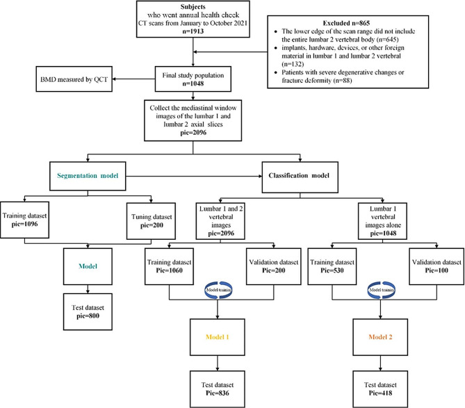
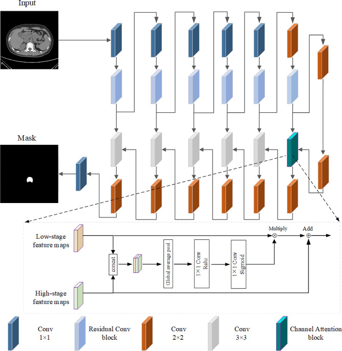
The chest CT images of 1048 health check subjects from January 2021 to June were retrospectively collected as the internal dataset (the segmentation model: 548 for training, 100 for tuning and 400 for test. The classification model: 530 for training, 100 for validation and 418 for test set). The subjects were divided into three categories according to the quantitative CT measurements, namely, normal, osteopenia and osteoporosis. First, a deep learning-based segmentation model was constructed, and the dice similarity coefficient(DSC) was used to compare the consistency between the model and manual labelling. Then, two classification models were established, namely, (i) model 1 (fusion feature construction of lumbar vertebral bodies 1 and 2) and (ii) model 2 (feature construction of lumbar 1 alone). Receiver operating characteristic curves were used to evaluate the diagnostic efficacy of the models, and the Delong test was used to compare the areas under the curve.
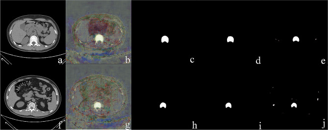
When the number of images in the training set was 300, the DSC value was 0.951 ± 0.030 in the test set. The results showed that the model 1 diagnosing normal, osteopenia and osteoporosis achieved an AUC of 0.990, 0.952 and 0.980; the model 2 diagnosing normal, osteopenia and osteoporosis achieved an AUC of 0.983, 0.940 and 0.978. The Delong test showed that there was no significant difference in area under the curve (AUC) values between the osteopenia group and osteoporosis group (P = 0.210, 0.546), while the AUC value of normal model 2 was higher than that of model 1 (0.990 vs. 0.983, P = 0.033).
This study proposed a chest CT deep learning model that achieves favorable performance on opportunistic osteoporosis screening using the lumbar 1 + lumbar 2 vertebral bodies fusion feature images. We further constructed the comparable model based on the lumbar 1 vertebra alone which can shorten the scan length, reduce the radiation dose received by patients, and reduce the training cost of technologists.
开发并评估一种基于胸部 CT 的深度学习模型,该模型通过融合腰椎 1+2 椎体特征图像,在机会性骨质疏松筛查方面取得了良好的性能,并探讨基于单个腰椎 1 椎体的模型的可行性和有效性。
回顾性收集了 2021 年 1 月至 6 月期间 1048 名健康体检者的胸部 CT 图像作为内部数据集(分割模型:548 个用于训练,100 个用于调整,400 个用于测试。分类模型:530 个用于训练,100 个用于验证,418 个用于测试集)。根据定量 CT 测量结果,将受试者分为三组,即正常、骨量减少和骨质疏松。首先,构建基于深度学习的分割模型,使用 Dice 相似系数(DSC)比较模型与手动标记之间的一致性。然后,建立两种分类模型,分别为(i)模型 1(腰椎 1 和 2 椎体融合特征构建)和(ii)模型 2(腰椎 1 单独特征构建)。使用受试者工作特征曲线评估模型的诊断效能,并使用 Delong 检验比较曲线下面积。
当训练集图像数量为 300 张时,测试集的 DSC 值为 0.951±0.030。结果表明,模型 1 诊断正常、骨量减少和骨质疏松的 AUC 值分别为 0.990、0.952 和 0.980;模型 2 诊断正常、骨量减少和骨质疏松的 AUC 值分别为 0.983、0.940 和 0.978。Delong 检验表明,骨量减少组和骨质疏松组的曲线下面积(AUC)值无显著差异(P=0.210,0.546),而正常模型 2 的 AUC 值高于模型 1(0.990 比 0.983,P=0.033)。
本研究提出了一种基于胸部 CT 的深度学习模型,该模型通过融合腰椎 1+2 椎体特征图像,在机会性骨质疏松筛查方面取得了良好的性能。我们进一步构建了基于单个腰椎 1 椎体的可比模型,该模型可以缩短扫描长度,降低患者接受的辐射剂量,并降低技术员的培训成本。