Department of Biomedical Engineering, Vanderbilt University, Nashville, TN, USA.
Department of Electrical and Computer Engineering, Vanderbilt University, Nashville, TN, USA.
Magn Reson Imaging. 2024 Jun;109:49-55. doi: 10.1016/j.mri.2024.02.016. Epub 2024 Feb 29.
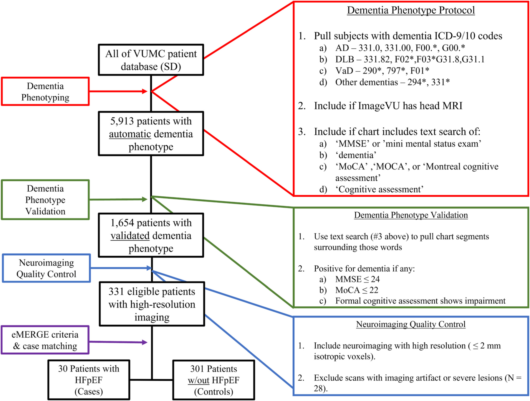
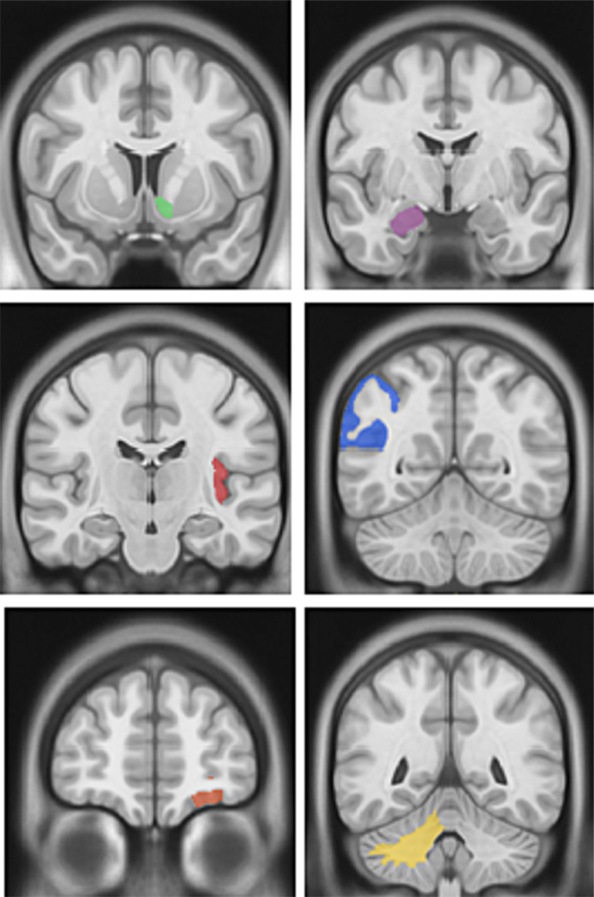
Heart failure with preserved ejection fraction (HFpEF) is an important, emerging risk factor for dementia, but it is not clear whether HFpEF contributes to a specific pattern of neuroanatomical changes in dementia. A major challenge to studying this is the relative paucity of datasets of patients with dementia, with/without HFpEF, and relevant neuroimaging. We sought to demonstrate the feasibility of using modern data mining tools to create and analyze clinical imaging datasets and identify the neuroanatomical signature of HFpEF-associated dementia. We leveraged the bioinformatics tools at Vanderbilt University Medical Center to identify patients with a diagnosis of dementia with and without comorbid HFpEF using the electronic health record. We identified high resolution, clinically-acquired neuroimaging data on 30 dementia patients with HFpEF (age 76.9 ± 8.12 years, 61% female) as well as 301 age- and sex-matched patients with dementia but without HFpEF to serve as comparators (age 76.2 ± 8.52 years, 60% female). We used automated image processing pipelines to parcellate the brain into 132 structures and quantify their volume. We found six regions with significant atrophy associated with HFpEF: accumbens area, amygdala, posterior insula, anterior orbital gyrus, angular gyrus, and cerebellar white matter. There were no regions with atrophy inversely associated with HFpEF. Patients with dementia and HFpEF have a distinct neuroimaging signature compared to patients with dementia only. Five of the six regions identified in are in the temporo-parietal region of the brain. Future studies should investigate mechanisms of injury associated with cerebrovascular disease leading to subsequent brain atrophy.
射血分数保留的心力衰竭(HFpEF)是痴呆的一个重要的新兴危险因素,但尚不清楚 HFpEF 是否导致痴呆的特定神经解剖变化模式。研究这一问题的主要挑战是,患有痴呆症、伴有/不伴有 HFpEF 的患者以及相关神经影像学的数据集相对较少。我们试图证明使用现代数据挖掘工具创建和分析临床成像数据集并确定与 HFpEF 相关的痴呆的神经解剖特征的可行性。我们利用范德比尔特大学医学中心的生物信息学工具,使用电子健康记录来识别患有痴呆症和合并 HFpEF 的患者和不伴有 HFpEF 的患者。我们确定了 30 名 HFpEF 痴呆症患者(年龄 76.9 ± 8.12 岁,61%为女性)以及 301 名年龄和性别匹配的无 HFpEF 痴呆症患者的高分辨率临床获得的神经影像学数据,用作对照(年龄 76.2 ± 8.52 岁,60%为女性)。我们使用自动图像处理管道将大脑分割成 132 个结构并量化它们的体积。我们发现与 HFpEF 相关的六个区域存在明显的萎缩:伏隔核区、杏仁核、后岛叶、前眶回、角回和小脑白质。没有与 HFpEF 相反的萎缩区域。与仅患有痴呆症的患者相比,患有痴呆症和 HFpEF 的患者具有明显的神经影像学特征。在鉴定出的六个区域中,有五个位于大脑的颞顶叶区域。未来的研究应该调查与导致随后脑萎缩的脑血管疾病相关的损伤机制。