Liu Yifan, Sun Bojian, Lin Yuan, Deng Hong, Wang Xinyi, Xu Chuanhao, Wang Kaibo, Yu Nan, Liu Rongqing, Han Mei
Department of Pathogenic Biology and Immunology, School of Basic Medical Sciences, Ningxia Medical University, Ningxia, P.R. China.
Department of Rheumatology and Immunology, The First Hospital of Hebei Medical University, Shijiazhuang, Hebei, P.R. China.
J Cancer. 2024 Feb 4;15(7):1816-1825. doi: 10.7150/jca.92192. eCollection 2024.
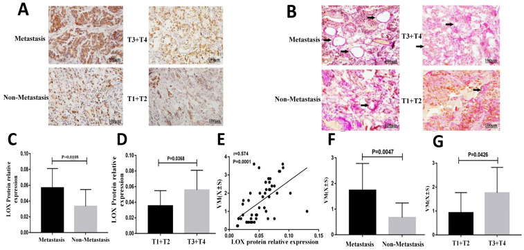
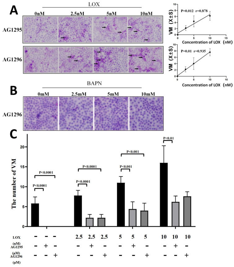
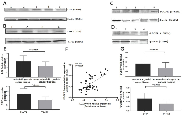
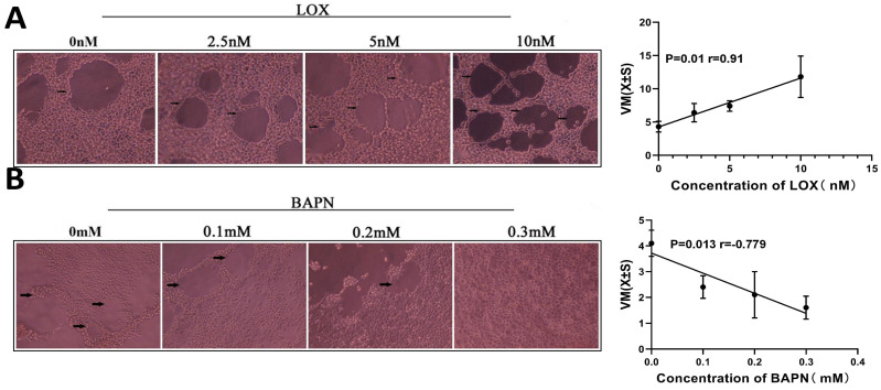
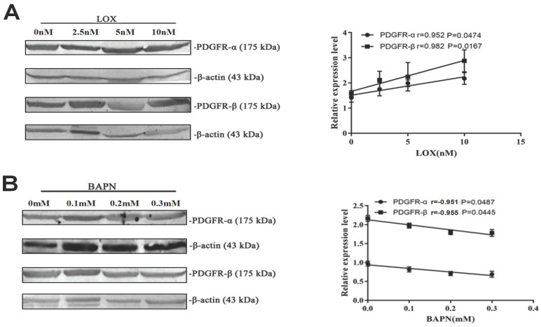
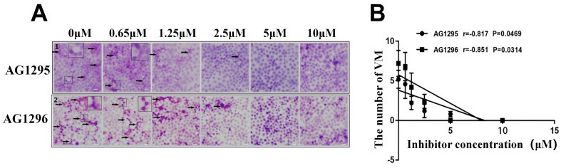
Vasculogenic mimicry (VM) generates an important supplementary form of blood supply in cancer, which many factors regulate. However, the effect of lysyl oxidase (LOX) on VM formation is unclear. In this study, gastric cancer tissues and cells were used to investigate the role of LOX in the formation of VM. The samples were collected from 49 patients with a final diagnosis of gastric cancer. According to metastasis (including lymph node metastases and distant metastases), gastric cancer samples were divided into metastasis and non-metastasis groups. Based on the degree of invasion, gastric cancer specimens were divided into T1 + T2 and T3 + T4 groups. The relative expression of LOX was detected using Western blot. The formation of VM was measured by double staining with CD34 and Periodic acid-Schiff (PAS) in gastric cancer tissue slices, and the correlation between LOX and VM was analyzed with Pearson's correlation analysis. Gastric cancer cell line BGC-803 was treated with LOX, β-aminopropionitrile (BAPN, an inhibitor of LOX), and AG1295 or AG1296 (inhibitors of the platelet-derived growth factor receptor). The formation of VM was then measured using PAS staining. The expression of platelet-derived growth factor receptor (PDGFR)α and PDGFRβ in gastric cancer cells was detected by Western blot. In gastric cancer samples, the level of LOX was higher in the metastasis group than in the non-metastasis group ( < 0.05) and in the T3 + T4 group than in the T1 + T2 group ( < 0.05). VM formation was greater in the T3+T4 group than in the T1+T2 group ( < 0.05) and in the metastasis group than in the non-metastasis group ( < 0.05). The expression level of LOX was positively correlated with VM formation ( < 0.01). In gastric cancer cells, LOX concentration was positively correlated with the degree of VM, and BAPN concentration was negatively correlated with the degree of VM ( <0.05). PDGFR levels in the T3+T4 and metastasis groups were relatively higher ( <0.01) and positively correlated with LOX levels in gastric cancer specimens ( < 0.01) The relative expression of PDGFRα and PDGFRβ in gastric cancer cells was up-regulated with increasing LOX and downregulated with increasing BAPN ( < 0.05). With inhibition of PDGFRα and PDGFRβ using AG1295 or AG1296, VM formation in gastric cancer cells decreased ( <0.05), but the number of VM structures increased while LOX was added ( < 0.05). LOX partially promotes the formation of VM in gastric cancer through the PDGF-PDGFR signaling pathway.
血管生成拟态(VM)在癌症中产生一种重要的补充性血液供应形式,受多种因素调控。然而,赖氨酰氧化酶(LOX)对VM形成的影响尚不清楚。本研究利用胃癌组织和细胞来探究LOX在VM形成中的作用。样本取自49例最终诊断为胃癌的患者。根据转移情况(包括淋巴结转移和远处转移),将胃癌样本分为转移组和非转移组。根据侵袭程度,将胃癌标本分为T1 + T2组和T3 + T4组。采用蛋白质免疫印迹法检测LOX的相对表达。通过对胃癌组织切片进行CD34和过碘酸希夫(PAS)双重染色来测定VM的形成,并采用Pearson相关性分析来分析LOX与VM之间的相关性。用LOX、β-氨基丙腈(BAPN,一种LOX抑制剂)以及AG1295或AG1296(血小板衍生生长因子受体抑制剂)处理胃癌细胞系BGC-803。然后采用PAS染色来测定VM的形成。通过蛋白质免疫印迹法检测胃癌细胞中血小板衍生生长因子受体(PDGFR)α和PDGFRβ的表达。在胃癌样本中,转移组的LOX水平高于非转移组(<0.05),T3 + T4组高于T1 + T2组(<0.05)。T3 + T4组的VM形成多于T1 + T2组(<0.05),转移组多于非转移组(<0.05)。LOX的表达水平与VM形成呈正相关(<0.01)。在胃癌细胞中,LOX浓度与VM程度呈正相关,BAPN浓度与VM程度呈负相关(<0.05)。T3 + T4组和转移组中的PDGFR水平相对较高(<0.01),且与胃癌标本中的LOX水平呈正相关(<0.01)。随着LOX增加,胃癌细胞中PDGFRα和PDGFRβ的相对表达上调,随着BAPN增加则下调(<0.05)。使用AG1295或AG1296抑制PDGFRα和PDGFRβ后,胃癌细胞中的VM形成减少(<0.05),但添加LOX时VM结构数量增加(<0.05)。LOX通过PDGF-PDGFR信号通路部分促进胃癌中VM的形成。