• 文献检索
  • 文档翻译
  • 深度研究
  • 学术资讯
  • Suppr Zotero 插件Zotero 插件
  • 邀请有礼
  • 套餐&价格
  • 历史记录
应用&插件
Suppr Zotero 插件Zotero 插件浏览器插件Mac 客户端Windows 客户端微信小程序
定价
高级版会员购买积分包购买API积分包
服务
文献检索文档翻译深度研究API 文档MCP 服务
关于我们
关于 Suppr公司介绍联系我们用户协议隐私条款
关注我们

Suppr 超能文献

核心技术专利:CN118964589B侵权必究
粤ICP备2023148730 号-1Suppr @ 2026

文献检索

告别复杂PubMed语法,用中文像聊天一样搜索,搜遍4000万医学文献。AI智能推荐,让科研检索更轻松。

立即免费搜索

文件翻译

保留排版,准确专业,支持PDF/Word/PPT等文件格式,支持 12+语言互译。

免费翻译文档

深度研究

AI帮你快速写综述,25分钟生成高质量综述,智能提取关键信息,辅助科研写作。

立即免费体验

计算病理学模型评估肾移植活检中肾小管间质腔的急性和慢性转化。

Computational pathology model to assess acute and chronic transformations of the tubulointerstitial compartment in renal allograft biopsies.

机构信息

Department of Pathology and Forensic Medicine, Institute of Biomedical Sciences of the Faculty of Medicine, Vilnius University, M. K. Ciurlionio Str. 21, 03101, Vilnius, Lithuania.

National Centre of Pathology, Vilnius University Hospital Santaros Klinikos, P. Baublio Str. 5, 08406, Vilnius, Lithuania.

出版信息

Sci Rep. 2024 Mar 4;14(1):5345. doi: 10.1038/s41598-024-55936-3.

DOI:10.1038/s41598-024-55936-3
PMID:38438513
原文链接:https://pmc.ncbi.nlm.nih.gov/articles/PMC10912734/
Abstract

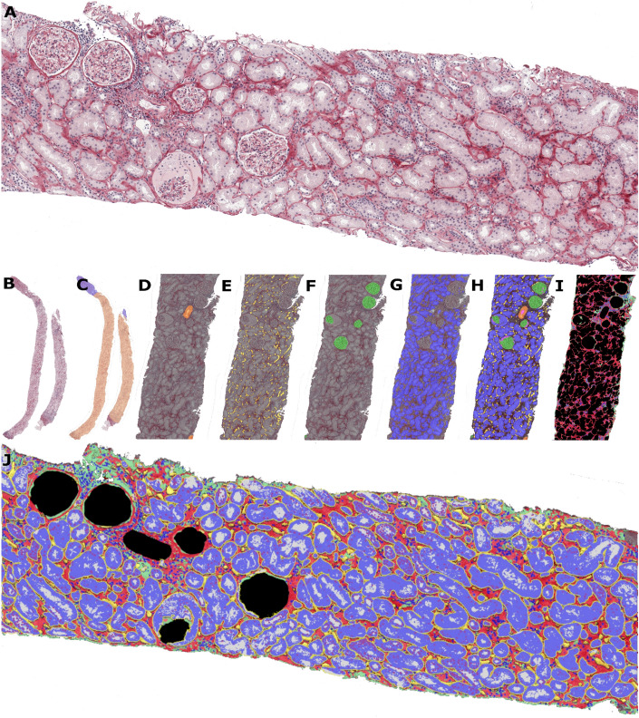
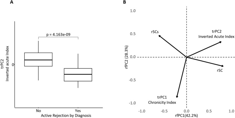
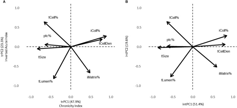
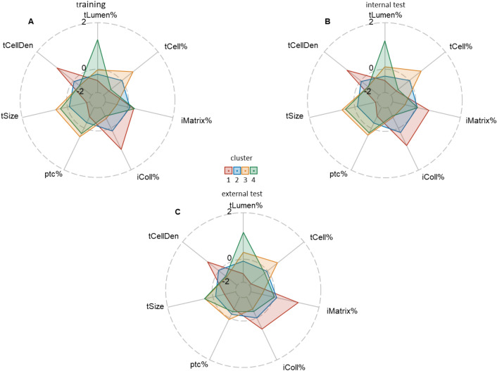
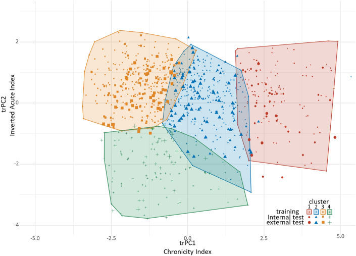
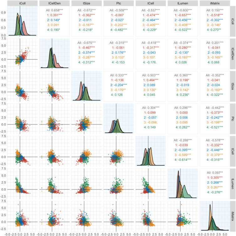
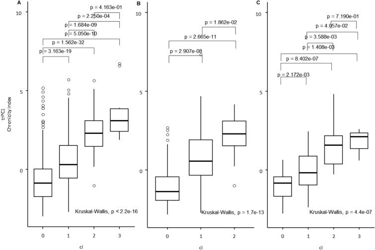
Managing patients with kidney allografts largely depends on biopsy diagnosis which is based on semiquantitative assessments of rejection features and extent of acute and chronic changes within the renal parenchyma. Current methods lack reproducibility while digital image data-driven computational models enable comprehensive and quantitative assays. In this study we aimed to develop a computational method for automated assessment of histopathology transformations within the tubulointerstitial compartment of the renal cortex. Whole slide images of modified Picrosirius red-stained biopsy slides were used for the training (n = 852) and both internal (n = 172) and external (n = 94) tests datasets. The pipeline utilizes deep learning segmentations of renal tubules, interstitium, and peritubular capillaries from which morphometry features were extracted. Seven indicators were selected for exploring the intrinsic spatial interactions within the tubulointerstitial compartment. A principal component analysis revealed two independent factors which can be interpreted as representing chronic and acute tubulointerstitial injury. A K-means clustering classified biopsies according to potential phenotypes of combined acute and chronic transformations of various degrees. We conclude that multivariate analyses of tubulointerstitial morphometry transformations enable extraction of and quantification of acute and chronic components of injury. The method is developed for renal allograft biopsies; however, the principle can be applied more broadly for kidney pathology assessment.

摘要

管理肾移植患者主要依赖于活检诊断,其基于对排斥特征的半定量评估以及肾实质内急性和慢性变化的程度。当前的方法缺乏可重复性,而数字图像数据驱动的计算模型则可以实现全面和定量的检测。在这项研究中,我们旨在开发一种用于自动评估肾皮质小管间区组织病理学转化的计算方法。使用改良的苦味酸天狼星红染色活检切片的全幻灯片图像进行训练(n=852)和内部(n=172)以及外部(n=94)测试数据集。该流水线利用深度学习对肾小管、间质和肾小管周毛细血管进行分割,从中提取形态计量学特征。选择了七个指标来探索小管间区内在的空间相互作用。主成分分析揭示了两个独立的因素,可解释为代表慢性和急性小管间损伤。K-均值聚类根据不同程度的急性和慢性联合转化的潜在表型对活检进行分类。我们得出结论,小管间形态计量学转化的多元分析可以提取和量化损伤的急性和慢性成分。该方法是为肾移植活检开发的;然而,该原理可以更广泛地应用于肾脏病理学评估。

https://cdn.ncbi.nlm.nih.gov/pmc/blobs/f71a/10912734/9d86ff6122dc/41598_2024_55936_Fig7_HTML.jpg
https://cdn.ncbi.nlm.nih.gov/pmc/blobs/f71a/10912734/b2eb44b7f453/41598_2024_55936_Fig1_HTML.jpg
https://cdn.ncbi.nlm.nih.gov/pmc/blobs/f71a/10912734/83207dfd67c5/41598_2024_55936_Fig2_HTML.jpg
https://cdn.ncbi.nlm.nih.gov/pmc/blobs/f71a/10912734/27c2cfccf35a/41598_2024_55936_Fig3_HTML.jpg
https://cdn.ncbi.nlm.nih.gov/pmc/blobs/f71a/10912734/23817ae2653f/41598_2024_55936_Fig4_HTML.jpg
https://cdn.ncbi.nlm.nih.gov/pmc/blobs/f71a/10912734/15e8ecd6e929/41598_2024_55936_Fig5_HTML.jpg
https://cdn.ncbi.nlm.nih.gov/pmc/blobs/f71a/10912734/faf6ce3a1226/41598_2024_55936_Fig6_HTML.jpg
https://cdn.ncbi.nlm.nih.gov/pmc/blobs/f71a/10912734/9d86ff6122dc/41598_2024_55936_Fig7_HTML.jpg

相似文献

1
Computational pathology model to assess acute and chronic transformations of the tubulointerstitial compartment in renal allograft biopsies.计算病理学模型评估肾移植活检中肾小管间质腔的急性和慢性转化。
Sci Rep. 2024 Mar 4;14(1):5345. doi: 10.1038/s41598-024-55936-3.
2
Evaluating tubulointerstitial compartments in renal biopsy specimens using a deep learning-based approach for classifying normal and abnormal tubules.使用深度学习方法评估肾活检标本中的肾小管间质隔室,用于分类正常和异常肾小管。
PLoS One. 2022 Jul 11;17(7):e0271161. doi: 10.1371/journal.pone.0271161. eCollection 2022.
3
Acute humoral rejection in kidney transplantation: II. Morphology, immunopathology, and pathologic classification.肾移植中的急性体液排斥反应:II. 形态学、免疫病理学及病理分类
J Am Soc Nephrol. 2002 Mar;13(3):779-787. doi: 10.1681/ASN.V133779.
4
Development of CD3 cell quantitation algorithms for renal allograft biopsy rejection assessment utilizing open source image analysis software.利用开源图像分析软件开发用于肾移植活检排斥评估的CD3细胞定量算法。
Virchows Arch. 2018 Feb;472(2):259-269. doi: 10.1007/s00428-017-2260-6. Epub 2017 Nov 8.
5
Data-Driven Chronic Allograft Phenotypes: A Novel and Validated Complement for Histologic Assessment of Kidney Transplant Biopsies.数据驱动的慢性移植物表型:一种新颖且经过验证的补充方法,用于评估肾移植活检的组织学表现。
J Am Soc Nephrol. 2022 Nov;33(11):2026-2039. doi: 10.1681/ASN.2022030290. Epub 2022 Sep 7.
6
Artificial intelligence and algorithmic computational pathology: an introduction with renal allograft examples.人工智能和算法计算病理学:以肾移植为例的介绍。
Histopathology. 2021 May;78(6):791-804. doi: 10.1111/his.14304. Epub 2021 Mar 8.
7
Expression of the chemokine receptor CCR1 in human renal allografts.趋化因子受体CCR1在人同种异体肾移植中的表达。
Nephrol Dial Transplant. 2007 Jun;22(6):1720-9. doi: 10.1093/ndt/gfm007. Epub 2007 Feb 13.
8
Deep learning-based classification of kidney transplant pathology: a retrospective, multicentre, proof-of-concept study.基于深度学习的肾移植病理学分类:一项回顾性、多中心、概念验证研究。
Lancet Digit Health. 2022 Jan;4(1):e18-e26. doi: 10.1016/S2589-7500(21)00211-9. Epub 2021 Nov 15.
9
Digital pathology evaluation of complement C4d component deposition in the kidney allograft biopsies is a useful tool to improve reproducibility of the scoring.数字化病理学评估肾移植活检中补体 C4d 成分的沉积是提高评分重复性的有用工具。
Diagn Pathol. 2011 Mar 30;6 Suppl 1(Suppl 1):S5. doi: 10.1186/1746-1596-6-S1-S5.
10
An evaluation of the Banff classification of early renal allograft biopsies and correlation with outcome.早期肾移植活检的Banff分类评估及其与预后的相关性。
Nephrol Dial Transplant. 1999 Oct;14(10):2364-9. doi: 10.1093/ndt/14.10.2364.

引用本文的文献

1
Artificial intelligence-enhanced interpretation of kidney transplant biopsy: focus on rejection.人工智能增强的肾移植活检解读:聚焦于排斥反应。
Curr Opin Organ Transplant. 2025 Jun 1;30(3):201-207. doi: 10.1097/MOT.0000000000001213. Epub 2025 Apr 1.

本文引用的文献

1
Unsatisfactory reproducibility of interstitial inflammation scoring in allograft kidney biopsy.移植肾活检中间质炎症评分的重复性不佳。
Sci Rep. 2023 May 1;13(1):7095. doi: 10.1038/s41598-023-33908-3.
2
Next-Generation Morphometry for pathomics-data mining in histopathology.下一代形态计量学在组织病理学病理组学数据挖掘中的应用。
Nat Commun. 2023 Jan 28;14(1):470. doi: 10.1038/s41467-023-36173-0.
3
Data-Driven Chronic Allograft Phenotypes: A Novel and Validated Complement for Histologic Assessment of Kidney Transplant Biopsies.
数据驱动的慢性移植物表型:一种新颖且经过验证的补充方法,用于评估肾移植活检的组织学表现。
J Am Soc Nephrol. 2022 Nov;33(11):2026-2039. doi: 10.1681/ASN.2022030290. Epub 2022 Sep 7.
4
Histological diagnosis from kidney transplant biopsy can contribute to prediction of graft survival.肾移植活检的组织学诊断有助于预测移植物的存活率。
Nephrology (Carlton). 2022 Jun;27(6):528-536. doi: 10.1111/nep.14028. Epub 2022 Feb 24.
5
Deep learning segmentation of glomeruli on kidney donor frozen sections.肾供体冰冻切片上肾小球的深度学习分割
J Med Imaging (Bellingham). 2021 Nov;8(6):067501. doi: 10.1117/1.JMI.8.6.067501. Epub 2021 Dec 20.
6
Thirty years of the International Banff Classification for Allograft Pathology: the past, present, and future of kidney transplant diagnostics.国际移植肾病理学班夫分类三十年:肾移植诊断的过去、现在与未来
Kidney Int. 2022 Apr;101(4):678-691. doi: 10.1016/j.kint.2021.11.028. Epub 2021 Dec 17.
7
Deep learning-based classification of kidney transplant pathology: a retrospective, multicentre, proof-of-concept study.基于深度学习的肾移植病理学分类:一项回顾性、多中心、概念验证研究。
Lancet Digit Health. 2022 Jan;4(1):e18-e26. doi: 10.1016/S2589-7500(21)00211-9. Epub 2021 Nov 15.
8
Deep learning in cancer diagnosis, prognosis and treatment selection.深度学习在癌症诊断、预后和治疗选择中的应用。
Genome Med. 2021 Sep 27;13(1):152. doi: 10.1186/s13073-021-00968-x.
9
Image Analysis Pipeline for Renal Allograft Evaluation and Fibrosis Quantification.用于肾移植评估和纤维化定量的图像分析流程
Kidney Int Rep. 2021 Apr 24;6(7):1878-1887. doi: 10.1016/j.ekir.2021.04.019. eCollection 2021 Jul.
10
Deep learning in histopathology: the path to the clinic.深度学习在组织病理学中的应用:通往临床的道路。
Nat Med. 2021 May;27(5):775-784. doi: 10.1038/s41591-021-01343-4. Epub 2021 May 14.