The University of Edinburgh Centre for Cardiovascular Science, University of Edinburgh, Room SU.305, Chancellor's Building, 49 Little France Crescent, Edinburgh, EH16 4SB, UK.
Department of Medical Physics, NHS Lothian, Royal Infirmary of Edinburgh, Edinburgh, UK.
Eur J Nucl Med Mol Imaging. 2024 Jul;51(8):2260-2270. doi: 10.1007/s00259-024-06670-5. Epub 2024 Mar 8.
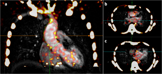
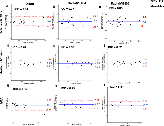
Non-invasive detection of pathological changes in thoracic aortic disease remains an unmet clinical need particularly for patients with congenital heart disease. Positron emission tomography combined with magnetic resonance imaging (PET-MRI) could provide a valuable low-radiation method of aortic surveillance in high-risk groups. Quantification of aortic microcalcification activity using sodium [F]fluoride holds promise in the assessment of thoracic aortopathies. We sought to evaluate aortic sodium [F]fluoride uptake in PET-MRI using three methods of attenuation correction compared to positron emission tomography computed tomography (PET-CT) in patients with bicuspid aortic valve, METHODS: Thirty asymptomatic patients under surveillance for bicuspid aortic valve disease underwent sodium [F]fluoride PET-CT and PET-MRI of the ascending thoracic aorta during a single visit. PET-MRI data were reconstructed using three iterations of attenuation correction (Dixon, radial gradient recalled echo with two [RadialVIBE-2] or four [RadialVIBE-4] tissue segmentation). Images were qualitatively and quantitatively analysed for aortic sodium [F]fluoride uptake on PET-CT and PET-MRI.
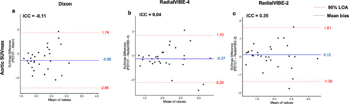
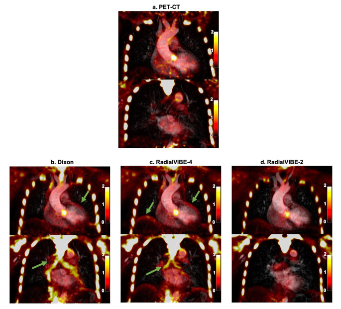
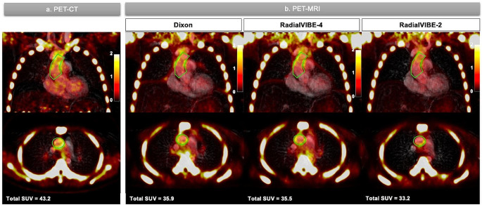
Aortic sodium [F]fluoride uptake on PET-MRI was visually comparable with PET-CT using each reconstruction and total aortic standardised uptake values on PET-CT strongly correlated with each PET-MRI attenuation correction method (Dixon R = 0.70; RadialVIBE-2 R = 0.63; RadialVIBE-4 R = 0.64; p < 0.001 for all). Breathing related artefact between soft tissue and lung were detected using Dixon and RadialVIBE-4 but not RadialVIBE-2 reconstructions, with the presence of this artefact adjacent to the atria leading to variations in blood pool activity estimates. Consequently, quantitative agreements between radiotracer activity on PET-CT and PET-MRI were most consistent with RadialVIBE-2.
Ascending aortic microcalcification analysis in PET-MRI is feasible with comparable findings to PET-CT. RadialVIBE-2 tissue attenuation correction correlates best with the reference standard of PET-CT and is less susceptible to artefact. There remain challenges in segmenting tissue types in PET-MRI reconstructions, and improved attenuation correction methods are required.
在胸主动脉疾病中,非侵入性检测病理变化仍然是一个未满足的临床需求,特别是对于先天性心脏病患者。正电子发射断层扫描结合磁共振成像(PET-MRI)可能为高危人群的主动脉监测提供一种有价值的低辐射方法。使用[F]氟酸钠定量主动脉微钙化活性有望评估胸主动脉病变。我们试图通过三种衰减校正方法在 PET-MRI 中评估主动脉[F]氟酸钠摄取,并与双瓣主动脉瓣患者的正电子发射断层扫描计算机断层扫描(PET-CT)进行比较。
30 例无症状的双瓣主动脉瓣疾病监测患者在一次就诊期间接受了[F]氟酸钠 PET-CT 和升主动脉的 PET-MRI 检查。使用三种迭代衰减校正(Dixon、具有两个[RadialVIBE-2]或四个[RadialVIBE-4]组织分割的径向梯度回波)重建 PET-MRI 数据。对 PET-CT 和 PET-MRI 上主动脉[F]氟酸钠摄取进行定性和定量分析。
使用每种重建方法,PET-MRI 上主动脉[F]氟酸钠摄取在视觉上与 PET-CT 相当,并且 PET-CT 上的总主动脉标准化摄取值与每种 PET-MRI 衰减校正方法均具有强烈相关性(Dixon R=0.70;RadialVIBE-2 R=0.63;RadialVIBE-4 R=0.64;p<0.001 均为)。使用 Dixon 和 RadialVIBE-4 可以检测到软组织和肺之间与呼吸相关的伪影,但 RadialVIBE-2 重建则不能,这种伪影位于心房附近,导致血池活性估计值发生变化。因此,与 PET-CT 相比,放射性示踪剂活性的定量一致性在 RadialVIBE-2 上最为一致。
在 PET-MRI 中进行升主动脉微钙化分析是可行的,其结果与 PET-CT 相似。RadialVIBE-2 组织衰减校正与参考标准 PET-CT 相关性最佳,并且不易受到伪影的影响。在 PET-MRI 重建中,仍然存在组织类型分割的挑战,需要改进衰减校正方法。