• 文献检索
  • 文档翻译
  • 深度研究
  • 学术资讯
  • Suppr Zotero 插件Zotero 插件
  • 邀请有礼
  • 套餐&价格
  • 历史记录
应用&插件
Suppr Zotero 插件Zotero 插件浏览器插件Mac 客户端Windows 客户端微信小程序
定价
高级版会员购买积分包购买API积分包
服务
文献检索文档翻译深度研究API 文档MCP 服务
关于我们
关于 Suppr公司介绍联系我们用户协议隐私条款
关注我们

Suppr 超能文献

核心技术专利:CN118964589B侵权必究
粤ICP备2023148730 号-1Suppr @ 2026

文献检索

告别复杂PubMed语法,用中文像聊天一样搜索,搜遍4000万医学文献。AI智能推荐,让科研检索更轻松。

立即免费搜索

文件翻译

保留排版,准确专业,支持PDF/Word/PPT等文件格式,支持 12+语言互译。

免费翻译文档

深度研究

AI帮你快速写综述,25分钟生成高质量综述,智能提取关键信息,辅助科研写作。

立即免费体验

通过扫频源光学相干断层扫描血管造影术测量糖尿病视网膜病变患者的视网膜和脉络膜血管灌注及厚度

Retinal and Choroidal Vascular Perfusion and Thickness Measurement in Diabetic Retinopathy Patients by the Swept-Source Optical Coherence Tomography Angiography.

作者信息

Liu Tingting, Lin Wei, Shi Genggeng, Wang Wenqi, Feng Meng, Xie Xiao, Liu Tong, Zhou Qingjun

机构信息

Eye Hospital of Shandong First Medical University (Shandong Eye Hospital), Jinan, China.

State Key Laboratory Cultivation Base, Shandong Provincial Key Laboratory of Ophthalmology, Shandong Eye Institute, Qingdao, China.

出版信息

Front Med (Lausanne). 2022 Mar 18;9:786708. doi: 10.3389/fmed.2022.786708. eCollection 2022.

DOI:10.3389/fmed.2022.786708
PMID:35372401
原文链接:https://pmc.ncbi.nlm.nih.gov/articles/PMC8971655/
Abstract

PURPOSE

To observe the changes in retinal and choroidal microstructures in patients with different stages of diabetic retinopathy (DR) and to evaluate the vascular perfusion of retina and choroid retinal thickness, retinal and choroidal vessel density by the swept-source optical coherence tomography angiography (SS-OCTA).

METHODS

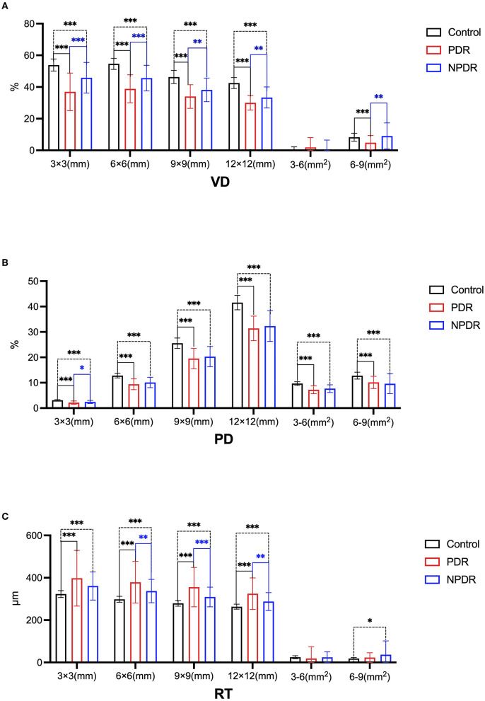
Subjects were divided into three groups: healthy control group (30 cases, 51 eyes), non-proliferative diabetic retinopathy (NPDR, 42 cases, 71 eyes) and proliferative diabetic retinopathy (PDR, 31 cases, 53 eyes). The area of the foveal avascular zone (FAZ), retinal and choroidal vascular perfusion, and the deep vascular complexes, including the intermediate capillary plexus (ICP) and deep capillary plexus (DCP) within the radius of 3, 6, 9, and 12 mm around the fovea were measured by SS-OCTA.

RESULTS

Compared with the healthy control group, DR patients presented significantly increased fovea avascular area, while vessel density (VD) in the ICP and DCP, vascular perfusion rate, and the retinal thickness were considerably decreased. There were significant differences in the retinal thickness, ICP, and DCP vessel densities between the control and NPDR groups, or control and PDR groups, or PDR and NPDR groups. The deep vascular perfusion rate also significantly differed between the control and PDR groups, but there was no significant difference between the PDR and NPDR groups. The choroidal perfusion exhibited significant differences across different areas and amongst the three groups. Furthermore, the choroidal thickness showed a significant difference between the PDR and NPDR groups.

CONCLUSION

Our results showed significant differences in the area of the avascular fovea and the VD of deep vascular complexes between DR patients and healthy control subjects. Moreover, there were significant differences in retinal VD, especially in the deep-retinalregions, choroidal perfusion, and the volume of large vessel choroid in DR patients with different degrees of disease severity. Notably, SS-OCTA provided in-depth information for detecting the potential VD damage in DR patients caused by a multitudeof factors.

摘要

目的

观察不同阶段糖尿病视网膜病变(DR)患者视网膜和脉络膜微观结构的变化,并通过扫频光学相干断层扫描血管造影(SS-OCTA)评估视网膜和脉络膜的血管灌注、视网膜厚度以及视网膜和脉络膜血管密度。

方法

将受试者分为三组:健康对照组(30例,51只眼)、非增殖性糖尿病视网膜病变组(NPDR,42例,71只眼)和增殖性糖尿病视网膜病变组(PDR,31例,53只眼)。通过SS-OCTA测量黄斑无血管区(FAZ)面积、视网膜和脉络膜血管灌注,以及黄斑周围3、6、9和12mm半径范围内的深层血管复合体,包括中间毛细血管丛(ICP)和深层毛细血管丛(DCP)。

结果

与健康对照组相比,DR患者的黄斑无血管区面积显著增加,而ICP和DCP中的血管密度(VD)、血管灌注率和视网膜厚度显著降低。对照组与NPDR组、对照组与PDR组或PDR组与NPDR组之间在视网膜厚度、ICP和DCP血管密度方面存在显著差异。对照组与PDR组之间的深层血管灌注率也有显著差异,但PDR组与NPDR组之间无显著差异。脉络膜灌注在不同区域和三组之间存在显著差异。此外,PDR组和NPDR组之间的脉络膜厚度存在显著差异。

结论

我们的结果显示,DR患者与健康对照者在无血管黄斑区面积和深层血管复合体的VD方面存在显著差异。此外,不同疾病严重程度的DR患者在视网膜VD,尤其是视网膜深层区域、脉络膜灌注和脉络膜大血管体积方面存在显著差异。值得注意的是,SS-OCTA为检测多种因素导致的DR患者潜在VD损伤提供了深入信息。

https://cdn.ncbi.nlm.nih.gov/pmc/blobs/5f29/8971655/8b8e66368d24/fmed-09-786708-g0001.jpg

相似文献

1
Retinal and Choroidal Vascular Perfusion and Thickness Measurement in Diabetic Retinopathy Patients by the Swept-Source Optical Coherence Tomography Angiography.通过扫频源光学相干断层扫描血管造影术测量糖尿病视网膜病变患者的视网膜和脉络膜血管灌注及厚度
Front Med (Lausanne). 2022 Mar 18;9:786708. doi: 10.3389/fmed.2022.786708. eCollection 2022.
2
Comparison of Chorioretinal Parameters in Diabetic Retinopathy with or without Pan-Retinal Photocoagulation Using Ultrawide-Field Swept-Source Optical Coherence Tomography Angiography.应用超广角扫频源光相干断层血管成像术比较伴有或不伴有全视网膜光凝的糖尿病性视网膜病变的脉络膜视网膜参数。
Ophthalmic Res. 2023;66(1):538-549. doi: 10.1159/000529335. Epub 2023 Jan 27.
3
Macular Capillary Perfusion in Chinese Patients With Diabetic Retinopathy Obtained With Optical Coherence Tomography Angiography.光学相干断层扫描血管造影术检测中国糖尿病视网膜病变患者的黄斑区毛细血管灌注情况
Ophthalmic Surg Lasers Imaging Retina. 2019 Apr 1;50(4):e88-e95. doi: 10.3928/23258160-20190401-12.
4
Retinal and choroidal microvascular characterization and density changes in different stages of diabetic retinopathy eyes.糖尿病视网膜病变不同阶段眼睛的视网膜和脉络膜微血管特征及密度变化
Front Med (Lausanne). 2023 Jul 25;10:1186098. doi: 10.3389/fmed.2023.1186098. eCollection 2023.
5
Correlation between vessel density and thickness in the retina and choroid of severe non-proliferative diabetic retinopathy patients.严重非增殖性糖尿病视网膜病变患者视网膜和脉络膜血管密度与厚度的相关性。
Front Endocrinol (Lausanne). 2024 May 14;15:1373363. doi: 10.3389/fendo.2024.1373363. eCollection 2024.
6
Analysis of retinal and choroidal characteristics in patients with early diabetic retinopathy using WSS-OCTA.应用 WSS-OCTA 分析早期糖尿病视网膜病变患者的视网膜和脉络膜特征。
Front Endocrinol (Lausanne). 2023 May 24;14:1184717. doi: 10.3389/fendo.2023.1184717. eCollection 2023.
7
Nonperfusion Area and Other Vascular Metrics by Wider Field Swept-Source OCT Angiography as Biomarkers of Diabetic Retinopathy Severity.广角扫频源光学相干断层扫描血管造影术测量的无灌注区及其他血管指标作为糖尿病视网膜病变严重程度的生物标志物
Ophthalmol Sci. 2022 Jun;2(2). doi: 10.1016/j.xops.2022.100144. Epub 2022 Mar 18.
8
Evaluating the Quantitative Foveal Avascular Zone and Retino-Choroidal Vessel Density Using Optical Coherence Tomography Angiography in a Healthy Indian Population.在健康印度人群中使用光学相干断层扫描血管造影术评估定量黄斑无血管区和视网膜脉络膜血管密度
Cureus. 2022 Aug 4;14(8):e27669. doi: 10.7759/cureus.27669. eCollection 2022 Aug.
9
Interaction Between the Distribution of Diabetic Retinopathy Lesions and the Association of Optical Coherence Tomography Angiography Scans With Diabetic Retinopathy Severity.糖尿病视网膜病变病变分布与光学相干断层扫描血管造影扫描与糖尿病视网膜病变严重程度的相关性分析。
JAMA Ophthalmol. 2020 Dec 1;138(12):1291-1297. doi: 10.1001/jamaophthalmol.2020.4516.
10
Correlations Between Optical Coherence Tomography Angiography Parameters and the Visual Acuity in Patients with Diabetic Retinopathy.糖尿病视网膜病变患者光学相干断层扫描血管造影参数与视力的相关性
Clin Ophthalmol. 2020 Apr 23;14:1107-1115. doi: 10.2147/OPTH.S248881. eCollection 2020.

引用本文的文献

1
Effects of Chinese Acupoint Eye Exercise on Ocular Biological Parameters.中医眼保健操对眼部生物学参数的影响。
Transl Vis Sci Technol. 2025 Jul 1;14(7):20. doi: 10.1167/tvst.14.7.20.
2
Retinal and choroidal blood flow changes in dialysis patients assessed by wide-field swept-source optical coherence tomography angiography.通过广角扫频源光学相干断层扫描血管造影评估透析患者的视网膜和脉络膜血流变化。
Front Med (Lausanne). 2025 May 14;12:1524503. doi: 10.3389/fmed.2025.1524503. eCollection 2025.
3
Diagnostic value of choroidal vascular density in predicting the progression of diabetic retinopathy.

本文引用的文献

1
Investigation of Macular Choroidal Thickness and Blood Flow Change by Optical Coherence Tomography Angiography After Posterior Scleral Reinforcement.后巩膜加固术后黄斑脉络膜厚度及血流变化的光学相干断层扫描血管造影研究
Front Med (Lausanne). 2021 Apr 29;8:658259. doi: 10.3389/fmed.2021.658259. eCollection 2021.
2
A Swept source optical coherence tomography angiography study: Imaging artifacts and comparison of non-perfusion areas with fluorescein angiography in diabetic macular edema.扫频源光相干断层扫描血管造影研究:糖尿病性黄斑水肿的成像伪影与荧光素血管造影的无灌注区比较。
PLoS One. 2021 Apr 8;16(4):e0249918. doi: 10.1371/journal.pone.0249918. eCollection 2021.
3
脉络膜血管密度在预测糖尿病视网膜病变进展中的诊断价值
Sci Rep. 2025 May 5;15(1):15671. doi: 10.1038/s41598-025-00528-y.
4
Three-Dimensional Choroidal Vessels Assessment in Diabetic Retinopathy.糖尿病视网膜病变中脉络膜血管的三维评估
Invest Ophthalmol Vis Sci. 2025 Mar 3;66(3):50. doi: 10.1167/iovs.66.3.50.
5
The role of optical coherence tomography angiography in assessing diabetic choroidopathy: a systematic review.光学相干断层扫描血管造影在评估糖尿病性脉络膜病变中的作用:一项系统评价。
Int J Retina Vitreous. 2025 Jan 31;11(1):10. doi: 10.1186/s40942-024-00618-5.
6
Evaluation of Retinal and Posterior Segment Vascular Changes Due to Systemic Hypoxia Using Optical Coherence Tomography Angiography.使用光学相干断层扫描血管造影术评估全身性缺氧引起的视网膜和眼后节血管变化
J Clin Med. 2024 Nov 7;13(22):6680. doi: 10.3390/jcm13226680.
7
Artificial Intelligence-Assisted Perfusion Density as Biomarker for Screening Diabetic Nephropathy.人工智能辅助灌注密度作为糖尿病肾病筛查的生物标志物。
Transl Vis Sci Technol. 2024 Oct 1;13(10):19. doi: 10.1167/tvst.13.10.19.
8
Correlation in retinal thickness and macular retina plus choroidal microcirculation in pediatric myopia.儿童近视的视网膜厚度与黄斑视网膜加脉络膜微循环的相关性。
Medicine (Baltimore). 2024 Jul 26;103(30):e38915. doi: 10.1097/MD.0000000000038915.
9
Advances in application of swept-source optical coherence tomography angiography in diagnosis and treatment of diabetic retinopathy.扫频源光学相干断层扫描血管造影术在糖尿病视网膜病变诊断和治疗中的应用进展
Front Ophthalmol (Lausanne). 2023 Feb 6;3:1116391. doi: 10.3389/fopht.2023.1116391. eCollection 2023.
10
Vascular changes of the choroid and their correlations with visual acuity in diabetic retinopathy.糖尿病视网膜病变中脉络膜血管变化及其与视力的相关性
Front Endocrinol (Lausanne). 2024 Feb 23;15:1327325. doi: 10.3389/fendo.2024.1327325. eCollection 2024.
Non-proliferative diabetic retinopathy.
非增殖性糖尿病视网膜病变
Dis Mon. 2021 May;67(5):101139. doi: 10.1016/j.disamonth.2021.101139. Epub 2021 Feb 1.
4
Detection of neovascularisation in the vitreoretinal interface slab using widefield swept-source optical coherence tomography angiography in diabetic retinopathy.在糖尿病性视网膜病变中,使用广角扫频源光学相干断层扫描血管造影术检测玻璃体视网膜界面层中的新生血管形成。
Br J Ophthalmol. 2022 Apr;106(4):534-539. doi: 10.1136/bjophthalmol-2020-317983. Epub 2020 Dec 21.
5
Comparison Between Graders in Detection of Diabetic Neovascularization With Swept Source Optical Coherence Tomography Angiography and Fluorescein Angiography.扫频源光学相干断层扫描血管造影术与荧光素血管造影术在糖尿病新生血管检测中分级者之间的比较
Am J Ophthalmol. 2021 Apr;224:292-300. doi: 10.1016/j.ajo.2020.11.020. Epub 2020 Dec 11.
6
Optical coherence tomography angiography in diabetic retinopathy: an updated review.光学相干断层扫描血管造影在糖尿病视网膜病变中的应用:最新综述。
Eye (Lond). 2021 Jan;35(1):149-161. doi: 10.1038/s41433-020-01233-y. Epub 2020 Oct 24.
7
Risk factors and prevalence of diabetic retinopathy: A protocol for meta-analysis.糖尿病视网膜病变的危险因素与患病率:一项荟萃分析方案
Medicine (Baltimore). 2020 Oct 16;99(42):e22695. doi: 10.1097/MD.0000000000022695.
8
Choroidal vascular changes in retinitis pigmentosa patients detected by optical coherence tomography angiography.通过光学相干断层扫描血管造影检测视网膜色素变性患者的脉络膜血管变化。
BMC Ophthalmol. 2020 Sep 29;20(1):384. doi: 10.1186/s12886-020-01640-5.
9
Macular vessel density in diabetes and diabetic retinopathy with swept-source optical coherence tomography angiography.用扫频源光相干断层扫描血管造影术观察糖尿病和糖尿病性视网膜病变的黄斑血管密度。
Graefes Arch Clin Exp Ophthalmol. 2020 Dec;258(12):2671-2679. doi: 10.1007/s00417-020-04832-3. Epub 2020 Jul 13.
10
Choroidal Thickness in Diabetes and Diabetic Retinopathy: A Swept Source OCT Study.脉络膜厚度在糖尿病和糖尿病性视网膜病变中的研究:扫频源光学相干断层扫描。
Invest Ophthalmol Vis Sci. 2020 Apr 9;61(4):29. doi: 10.1167/iovs.61.4.29.