Nakhjavani Maryam, Smith Eric, Yeo Kenny, Tomita Yoko, Price Timothy J, Yool Andrea, Townsend Amanda R, Hardingham Jennifer E
Molecular Oncology, Basil Hetzel Institute for Translational Health Research, The Queen Elizabeth Hospital, Woodville South, SA, Australia.
Adelaide Medical School, University of Adelaide, Adelaide, SA, Australia.
J Ginseng Res. 2024 Mar;48(2):171-180. doi: 10.1016/j.jgr.2021.05.008. Epub 2021 Jun 3.
Epimers of ginsenoside Rg3 (Rg3) have a low bioavailability and are prone to deglycosylation, which produces epimers of ginsenoside Rh2 (S-Rh2 and R-Rh2) and protopanaxadiol (S-PPD and R-PPD). The aim of this study was to compare the efficacy and potency of these molecules as anti-cancer agents.
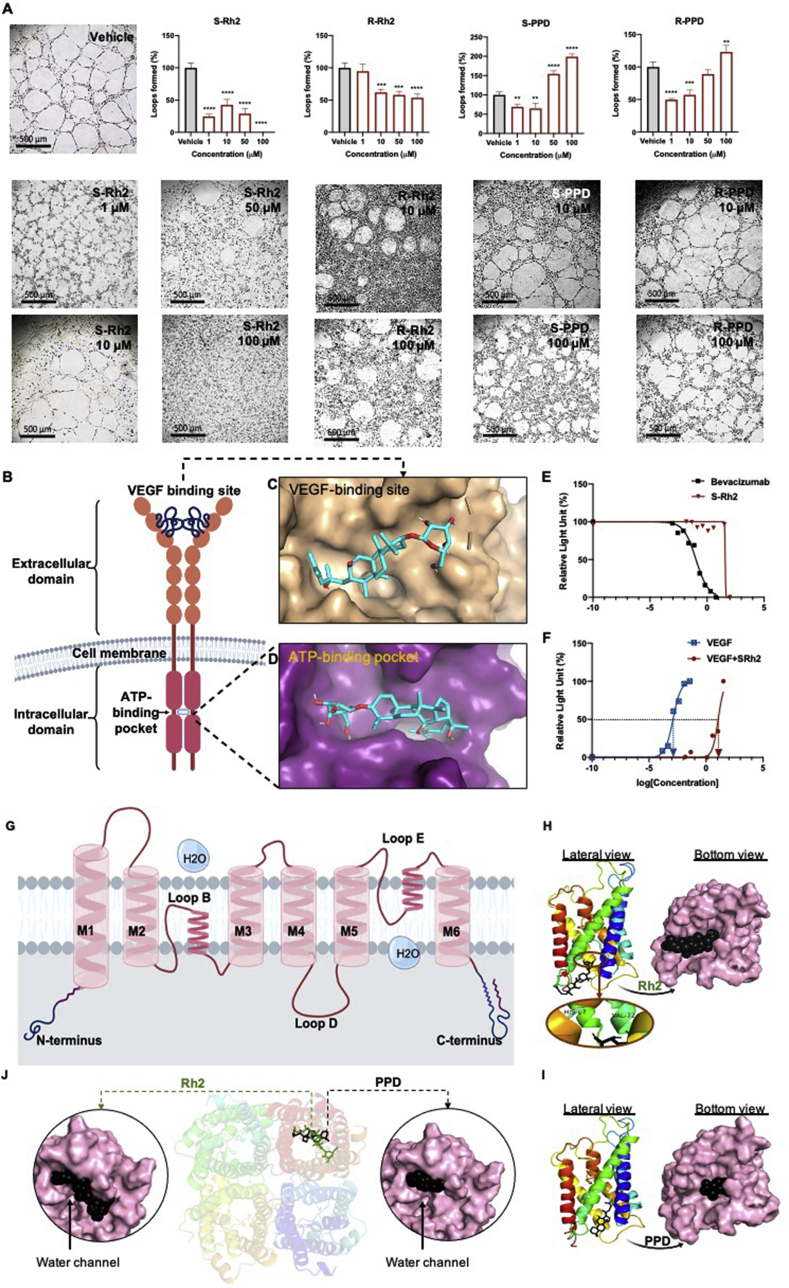
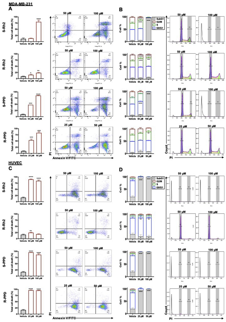
Crystal violet staining was used to study the anti-proliferatory action of the molecules on a human epithelial breast cancer cell line, MDA-MB-231, and human umbilical vein endothelial cells (HUVEC) and compare their potency. Cell death and cell cycle were studied using flow cytometry and mode of cell death was studied using live cell imaging. Anti-angiogenic effects of the drug were studied using loop formation assay. Molecular docking showed the interaction of these molecules with vascular endothelial growth factor receptor-2 (VEGFR2) and aquaporin (AQP) water channels. VEGF bioassay was used to study the interaction of Rh2 with VEGFR2, .
HUVEC was the more sensitive cell line to the anti-proliferative effects of S-Rh2, S-PPD and R-PPD. The molecules induced necroptosis/necrosis in MDA-MB-231 and apoptosis in HUVEC. S-Rh2 was the most potent inhibitor of loop formation. molecular docking predicted a good binding score between Rh2 or PPD and the ATP-binding pocket of VEGFR2. VEGF bioassay showed that Rh2 was an allosteric modulator of VEGFR2. In addition, SRh2 and PPD had good binding scores with AQP1 and AQP5, both of which play roles in cell migration and proliferation.
The combination of these molecules might be responsible for the anti-cancer effects observed by Rg3.
人参皂苷Rg3(Rg3)的差向异构体生物利用度低,且易于去糖基化,从而产生人参皂苷Rh2的差向异构体(S-Rh2和R-Rh2)以及原人参二醇(S-PPD和R-PPD)。本研究的目的是比较这些分子作为抗癌剂的疗效和效力。
采用结晶紫染色研究这些分子对人上皮性乳腺癌细胞系MDA-MB-231和人脐静脉内皮细胞(HUVEC)的抗增殖作用,并比较它们的效力。使用流式细胞术研究细胞死亡和细胞周期,使用活细胞成像研究细胞死亡模式。使用环形成试验研究该药物的抗血管生成作用。分子对接显示这些分子与血管内皮生长因子受体-2(VEGFR2)和水通道蛋白(AQP)水通道之间的相互作用。使用VEGF生物测定法研究Rh2与VEGFR2的相互作用。
HUVEC对S-Rh2、S-PPD和R-PPD的抗增殖作用更敏感。这些分子在MDA-MB-231中诱导坏死性凋亡/坏死,在HUVEC中诱导凋亡。S-Rh2是环形成的最有效抑制剂。分子对接预测Rh2或PPD与VEGFR2的ATP结合口袋之间具有良好的结合分数。VEGF生物测定表明Rh2是VEGFR2的变构调节剂。此外,SRh2和PPD与AQP1和AQP5具有良好的结合分数,这两者在细胞迁移和增殖中均起作用。
这些分子的组合可能是Rg3所观察到的抗癌作用的原因。