Reynoso-Mejia Carlos A, Troville Jonathan, Wagner Martin G, Hoppel Bernice, Lee Fred T, Szczykutowicz Timothy P
Department of Radiology, University of Wisconsin-Madison, Madison, WI, 53705, USA.
Department of Medical Physics, University of Wisconsin-Madison, Madison, WI, 53705, USA.
BMC Biomed Eng. 2024 Mar 11;6(1):2. doi: 10.1186/s42490-024-00076-y.
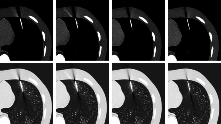
MAR algorithms have not been productized in interventional imaging because they are too time-consuming. Application of a beam hardening filter can mitigate metal artifacts and doesn't increase computational burden. We evaluate the ability to reduce metal artifacts of a 0.5 mm silver (Ag) additional filter in a Multidetector Computed Tomography (MDCT) scanner during CT-guided biopsy procedures.
A biopsy needle was positioned inside the lung field of an anthropomorphic phantom (Lungman, Kyoto Kagaku, Kyoto, Japan). CT acquisitions were performed with beam energies of 100 kV, 120 kV, 135 kV, and 120 kV with the Ag filter and reconstructed using a filtered back projection algorithm. For each measurement, the CTDIvol was kept constant at 1 mGy. Quantitative profiles placed in three regions of the artifact (needle, needle tip, and trajectory artifacts) were used to obtain metrics (FWHM, FWTM, width at - 100 HU, and absolute error in HU) to evaluate the blooming artifact, artifact width, change in CT number, and artifact range. An image quality analysis was carried out through image noise measurement. A one-way analysis of variance (ANOVA) test was used to find significant differences between the conventional CT beam energies and the Ag filtered 120 kV beam.
The 120 kV-Ag is shown to have the shortest range of artifacts compared to the other beam energies. For needle tip and trajectory artifacts, a significant reduction of - 53.6% (p < 0.001) and - 48.7% (p < 0.001) in the drop of the CT number was found, respectively, in comparison with the reference beam of 120 kV as well as a significant decrease of up to - 34.7% in the artifact width (width at - 100 HU, p < 0.001). Also, a significant reduction in the blooming artifact of - 14.2% (FWHM, p < 0.001) and - 53.3% (FWTM, p < 0.001) was found in the needle artifact. No significant changes (p > 0.05) in image noise between the conventional energies and the 120 kV-Ag were found.
A 0.5 mm Ag additional MDCT filter demonstrated consistent metal artifact reduction generated by the biopsy needle. This reduction may lead to a better depiction of the target and surrounding structures while maintaining image quality.
MAR算法尚未在介入成像中实现产品化,因为其耗时过长。应用束硬化滤波器可以减轻金属伪影,且不会增加计算负担。我们评估了在CT引导活检程序期间,多排螺旋CT(MDCT)扫描仪中0.5毫米银(Ag)附加滤波器减少金属伪影的能力。
将活检针置于仿真人体模型(Lungman,日本京都Kagaku公司,京都)的肺野内。使用100 kV、120 kV、135 kV和120 kV的束能量并带有Ag滤波器进行CT采集,并使用滤波反投影算法重建。对于每次测量,CTDIvol保持恒定在1 mGy。放置在伪影的三个区域(针、针尖和轨迹伪影)的定量剖面图用于获得指标(半高宽、十分位全宽、-100 HU处的宽度和HU中的绝对误差),以评估光晕伪影、伪影宽度、CT值变化和伪影范围。通过图像噪声测量进行图像质量分析。使用单因素方差分析(ANOVA)测试来发现传统CT束能量与Ag滤波的120 kV束之间的显著差异。
与其他束能量相比,120 kV-Ag显示出最短的伪影范围。对于针尖和轨迹伪影,与120 kV的参考束相比,CT值下降分别显著降低了-53.6%(p < 0.001)和-48.7%(p < 0.001),并且伪影宽度(-100 HU处的宽度)显著降低了高达-34.7%(p < 0.001)。此外,在针伪影中,光晕伪影分别显著降低了-14.2%(半高宽,p < 0.001)和-53.3%(十分位全宽,p < 0.001)。在传统能量与120 kV-Ag之间未发现图像噪声有显著变化(p > 0.05)。
0.5毫米Ag附加MDCT滤波器显示出由活检针产生的金属伪影持续减少。这种减少可能在保持图像质量的同时,更好地描绘目标和周围结构。