• 文献检索
  • 文档翻译
  • 深度研究
  • 学术资讯
  • Suppr Zotero 插件Zotero 插件
  • 邀请有礼
  • 套餐&价格
  • 历史记录
应用&插件
Suppr Zotero 插件Zotero 插件浏览器插件Mac 客户端Windows 客户端微信小程序
定价
高级版会员购买积分包购买API积分包
服务
文献检索文档翻译深度研究API 文档MCP 服务
关于我们
关于 Suppr公司介绍联系我们用户协议隐私条款
关注我们

Suppr 超能文献

核心技术专利:CN118964589B侵权必究
粤ICP备2023148730 号-1Suppr @ 2026

文献检索

告别复杂PubMed语法,用中文像聊天一样搜索,搜遍4000万医学文献。AI智能推荐,让科研检索更轻松。

立即免费搜索

文件翻译

保留排版,准确专业,支持PDF/Word/PPT等文件格式,支持 12+语言互译。

免费翻译文档

深度研究

AI帮你快速写综述,25分钟生成高质量综述,智能提取关键信息,辅助科研写作。

立即免费体验

掺杂有源自沙龙虾壳的羟基磷灰石的电纺纳米纤维支架用于骨组织工程。

Nanofibrous electrospun scaffold doped with hydroxyapatite derived from sand lobster shell () for bone tissue engineering.

作者信息

Dinatha I Kadek Hariscandra, Diputra Arian H, Wihadmadyatami Hevi, Partini Juliasih, Yusuf Yusril

机构信息

Department of Physics, Faculty of Mathematics and Natural Science, Universitas Gadjah Mada Yogyakarta Indonesia

Department of Anatomy, Faculty of Veterinary Medicine, Universitas Gadjah Mada Yogyakarta Indonesia.

出版信息

RSC Adv. 2024 Mar 11;14(12):8222-8239. doi: 10.1039/d4ra00619d. eCollection 2024 Mar 6.

DOI:10.1039/d4ra00619d
PMID:38469192
原文链接:https://pmc.ncbi.nlm.nih.gov/articles/PMC10925909/
Abstract

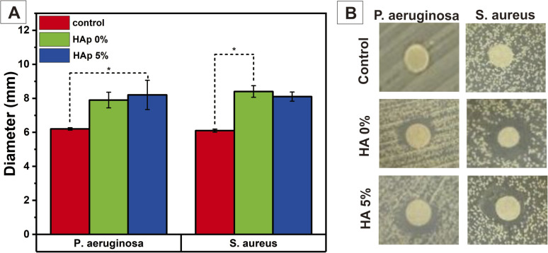
Healing of significant segmental bone defects remains a challenge, and various studies attempt to make materials that mimic bone structures and have biocompatibility, bioactivity, biodegradability, and osteoconductivity to native bone tissues. In this work, a nanofiber scaffold membrane of polyvinyl alcohol (PVA)/polyvinylpyrrolidone (PVP)/chitosan (CS) combined with hydroxyapatite (HAp) from sand lobster (SL; ) shells, as a calcium source, was successfully synthesized to mimic the nanoscale extracellular matrix (ECM) in the native bone. The HAp from SL shells was synthesized by co-precipitation method with Ca/P of 1.67 and incorporated into the nanofiber membrane PVA/PVP/CS synthesized by the electrospinning method with varying concentrations, 0, 1, 3, and 5% (w/v). Based on the morphological and physicochemical analysis, the addition of HAp into the nanofiber successfully showed incorporation into the nanofiber with small agglomeration at HAp concentrations of 1, 3, and 5% (w/v). This led to a smaller fiber diameter with higher concentration of Hap, and incorporating HAp into the nanofiber could improve the mechanical properties of the nanofiber closer to the trabecula bone. Moreover, in general, swelling due to water absorption increases due to higher hydrophilicity at higher HAp concentrations and leads to the improvement of the degradation process and protein adsorption of the nanofiber. Biomineralization in a simulated body fluid (SBF) solution confirms that the HAp in the nanofiber increases bioactivity, and it can be seen that more apatite is formed during longer immersion in the SBF solution. The nanofiber PVA/PVP/CS HAp 5% has the most potential for osteoblast (MC3T3E1) cell viability after being incubated for 24 h, and it allowed the cell to attach and proliferate. Additionally, the higher HAp concentration in the nanofiber scaffold membrane can significantly promote the osteogenic differentiation of MC3T3E1 cells. Overall, the PVA/PVP/CS/HAp 5% nanofiber scaffold membrane has the most potential for bone tissue engineering.

摘要

大面积节段性骨缺损的修复仍然是一项挑战,各种研究试图制造出模仿骨结构且具有生物相容性、生物活性、生物可降解性以及对天然骨组织具有骨传导性的材料。在本研究中,成功合成了一种由聚乙烯醇(PVA)/聚乙烯吡咯烷酮(PVP)/壳聚糖(CS)组成的纳米纤维支架膜,并结合了来自沙龙虾(SL)壳的羟基磷灰石(HAp)作为钙源,以模仿天然骨中的纳米级细胞外基质(ECM)。通过共沉淀法合成了Ca/P为1.67的SL壳HAp,并将其以0、1、3和5%(w/v)的不同浓度掺入通过电纺丝法合成的纳米纤维膜PVA/PVP/CS中。基于形态学和物理化学分析,在HAp浓度为1、3和5%(w/v)时,将HAp添加到纳米纤维中成功显示其掺入纳米纤维且团聚较小。这导致在较高Hap浓度下纤维直径更小,并且将HAp掺入纳米纤维可以改善纳米纤维的机械性能,使其更接近小梁骨。此外,总体而言,由于在较高HAp浓度下亲水性更高,吸水导致的肿胀增加,从而改善了纳米纤维的降解过程和蛋白质吸附。在模拟体液(SBF)溶液中的生物矿化证实了纳米纤维中的HAp增加了生物活性,并且可以看到在SBF溶液中浸泡更长时间时形成了更多的磷灰石。纳米纤维PVA/PVP/CS HAp 5%在孵育24小时后对成骨细胞(MC3T3E1)的细胞活力最具潜力,并且它允许细胞附着和增殖。此外,纳米纤维支架膜中较高的HAp浓度可以显著促进MC3T3E1细胞的成骨分化。总体而言,PVA/PVP/CS/HAp 5%纳米纤维支架膜在骨组织工程方面最具潜力。

https://cdn.ncbi.nlm.nih.gov/pmc/blobs/bd76/10925909/18694e71dfc4/d4ra00619d-f16.jpg
https://cdn.ncbi.nlm.nih.gov/pmc/blobs/bd76/10925909/e42bceedc021/d4ra00619d-f1.jpg
https://cdn.ncbi.nlm.nih.gov/pmc/blobs/bd76/10925909/ca25481d64a6/d4ra00619d-f2.jpg
https://cdn.ncbi.nlm.nih.gov/pmc/blobs/bd76/10925909/b99ad9705f8b/d4ra00619d-f3.jpg
https://cdn.ncbi.nlm.nih.gov/pmc/blobs/bd76/10925909/75a649ac568e/d4ra00619d-f4.jpg
https://cdn.ncbi.nlm.nih.gov/pmc/blobs/bd76/10925909/ebc557c35825/d4ra00619d-f5.jpg
https://cdn.ncbi.nlm.nih.gov/pmc/blobs/bd76/10925909/b90826f3700c/d4ra00619d-f6.jpg
https://cdn.ncbi.nlm.nih.gov/pmc/blobs/bd76/10925909/54373d826ff0/d4ra00619d-f7.jpg
https://cdn.ncbi.nlm.nih.gov/pmc/blobs/bd76/10925909/9f42920fadd4/d4ra00619d-f8.jpg
https://cdn.ncbi.nlm.nih.gov/pmc/blobs/bd76/10925909/5148d0fb34ab/d4ra00619d-f9.jpg
https://cdn.ncbi.nlm.nih.gov/pmc/blobs/bd76/10925909/9b9248d3047a/d4ra00619d-f10.jpg
https://cdn.ncbi.nlm.nih.gov/pmc/blobs/bd76/10925909/d279f4cf9fe0/d4ra00619d-f11.jpg
https://cdn.ncbi.nlm.nih.gov/pmc/blobs/bd76/10925909/087195f7616e/d4ra00619d-f12.jpg
https://cdn.ncbi.nlm.nih.gov/pmc/blobs/bd76/10925909/469e25234317/d4ra00619d-f13.jpg
https://cdn.ncbi.nlm.nih.gov/pmc/blobs/bd76/10925909/afe7a798e620/d4ra00619d-f14.jpg
https://cdn.ncbi.nlm.nih.gov/pmc/blobs/bd76/10925909/f13544d3cc21/d4ra00619d-f15.jpg
https://cdn.ncbi.nlm.nih.gov/pmc/blobs/bd76/10925909/18694e71dfc4/d4ra00619d-f16.jpg

相似文献

1
Nanofibrous electrospun scaffold doped with hydroxyapatite derived from sand lobster shell () for bone tissue engineering.掺杂有源自沙龙虾壳的羟基磷灰石的电纺纳米纤维支架用于骨组织工程。
RSC Adv. 2024 Mar 11;14(12):8222-8239. doi: 10.1039/d4ra00619d. eCollection 2024 Mar 6.
2
Porous scaffold hydroxyapatite from sand lobster shells (Panulirus homarus) using polyethylene oxide/chitosan as polymeric porogen for bone tissue engineering.以聚乙烯氧化物/壳聚糖为高分子成孔剂,从沙蟹(Panulirus homarus)壳中提取多孔支架羟基磷灰石,用于骨组织工程。
J Biomed Mater Res B Appl Biomater. 2024 Jan;112(1):e35341. doi: 10.1002/jbm.b.35341. Epub 2023 Oct 25.
3
Electrospun polyvinyl alcohol nanofiber scaffolds incorporated strontium-substituted hydroxyapatite from sand lobster shells: synthesis, characterization, and in vitro biological properties.静电纺丝聚乙烯醇纳米纤维支架结合了来自龙虾壳的锶取代羟基磷灰石:合成、表征及体外生物学特性
Biomed Mater. 2024 Sep 23. doi: 10.1088/1748-605X/ad7e92.
4
Nanofibrous poly(vinyl alcohol)/chitosan contained carbonated hydroxyapatite nanoparticles scaffold for bone tissue engineering.用于骨组织工程的含碳酸羟基磷灰石纳米粒子的纳米纤维聚(乙烯醇)/壳聚糖支架。
Mater Sci Eng C Mater Biol Appl. 2020 Feb;107:110347. doi: 10.1016/j.msec.2019.110347. Epub 2019 Oct 23.
5
Optimization of Biologically Inspired Electrospun Scaffold for Effective Use in Bone Regenerative Applications.用于骨再生应用的生物启发式电纺支架的优化,以实现有效利用。
Polymers (Basel). 2024 Jul 15;16(14):2023. doi: 10.3390/polym16142023.
6
Fibrous PVA Matrix Containing Strontium-Substituted Hydroxyapatite Nanoparticles from Golden Apple Snail ( L.) Shells for Bone Tissue Engineering.用于骨组织工程的含来自福寿螺(L.)贝壳的锶取代羟基磷灰石纳米颗粒的纤维状聚乙烯醇基质
Bioengineering (Basel). 2023 Jul 17;10(7):844. doi: 10.3390/bioengineering10070844.
7
Preparation and performance evaluation of chitosan/polyvinylpyrrolidone/polyvinyl alcohol electrospun nanofiber membrane for heavy metal ions and organic pollutants removal.壳聚糖/聚乙烯吡咯烷酮/聚乙烯醇静电纺丝纳米纤维膜的制备及其对重金属离子和有机污染物去除的性能评价。
Int J Biol Macromol. 2022 Jun 15;210:76-84. doi: 10.1016/j.ijbiomac.2022.05.017. Epub 2022 May 6.
8
Fabrication of Mesoporous Silica Nanoparticle-Incorporated Coaxial Nanofiber for Evaluating the In Vitro Osteogenic Potential.介孔硅纳米粒子复合同轴纳米纤维的制备及其体外成骨性能评价
Appl Biochem Biotechnol. 2022 Jan;194(1):302-322. doi: 10.1007/s12010-021-03741-3. Epub 2021 Nov 11.
9
Multifunctional electrospun nanofibrous scaffold enriched with alendronate and hydroxyapatite for balancing osteogenic and osteoclast activity to promote bone regeneration.富含阿仑膦酸盐和羟基磷灰石的多功能电纺纳米纤维支架,用于平衡成骨和破骨细胞活性以促进骨再生。
Front Bioeng Biotechnol. 2023 Nov 9;11:1302594. doi: 10.3389/fbioe.2023.1302594. eCollection 2023.
10
Functionalized cellulose nanofibrils in carbonate-substituted hydroxyapatite nanorod-based scaffold from long-spined sea urchin () shells reinforced with polyvinyl alcohol for alveolar bone tissue engineering.基于长棘海胆()壳的碳酸盐取代羟基磷灰石纳米棒支架中的功能化纤维素纳米纤维,用聚乙烯醇增强,用于牙槽骨组织工程。
RSC Adv. 2023 Nov 3;13(46):32444-32456. doi: 10.1039/d3ra06165e. eCollection 2023 Oct 31.

引用本文的文献

1
Dual-Layer Natamycin and Boric-Acid-Reinforced PVA/Chitosan by 3D Printing and Electrospinning Method: Characterization and In Vitro Evaluation.通过3D打印和静电纺丝法制备的双层纳他霉素和硼酸增强PVA/壳聚糖:表征与体外评价
Polymers (Basel). 2025 Jun 17;17(12):1673. doi: 10.3390/polym17121673.
2
Nano-Hydroxyapatite/Poly(methyl methacrylate) Composite Bone Scaffold: Surfactant Surface Effects.纳米羟基磷灰石/聚甲基丙烯酸甲酯复合骨支架:表面活性剂的表面效应
Polymers (Basel). 2025 Apr 23;17(9):1148. doi: 10.3390/polym17091148.
3
A comparative X-ray diffraction analysis of Srsubstituted hydroxyapatite from sand lobster shell waste using various methods.

本文引用的文献

1
Multifunctional electrospun nanofibrous scaffold enriched with alendronate and hydroxyapatite for balancing osteogenic and osteoclast activity to promote bone regeneration.富含阿仑膦酸盐和羟基磷灰石的多功能电纺纳米纤维支架,用于平衡成骨和破骨细胞活性以促进骨再生。
Front Bioeng Biotechnol. 2023 Nov 9;11:1302594. doi: 10.3389/fbioe.2023.1302594. eCollection 2023.
2
The enhanced properties and bioactivity of poly-ε-caprolactone/poly lactic--glycolic acid doped with carbonate hydroxyapatite-egg white.掺杂有碳酸羟基磷灰石-蛋清的聚ε-己内酯/聚乳酸-乙醇酸的增强性能和生物活性。
RSC Adv. 2023 Nov 24;13(49):34427-34438. doi: 10.1039/d3ra07486b. eCollection 2023 Nov 22.
3
使用各种方法对来自沙龙虾壳废料的锶取代羟基磷灰石进行的X射线衍射对比分析。
Heliyon. 2025 Jan 8;11(2):e41781. doi: 10.1016/j.heliyon.2025.e41781. eCollection 2025 Jan 30.
4
Polysaccharide Hydroxyapatite (Nano)composites and Their Biomedical Applications: An Overview of Recent Years.多糖羟基磷灰石(纳米)复合材料及其生物医学应用:近年来概述
ACS Omega. 2024 Jul 2;9(28):30035-30070. doi: 10.1021/acsomega.4c02170. eCollection 2024 Jul 16.
Porous scaffold hydroxyapatite from sand lobster shells (Panulirus homarus) using polyethylene oxide/chitosan as polymeric porogen for bone tissue engineering.
以聚乙烯氧化物/壳聚糖为高分子成孔剂,从沙蟹(Panulirus homarus)壳中提取多孔支架羟基磷灰石,用于骨组织工程。
J Biomed Mater Res B Appl Biomater. 2024 Jan;112(1):e35341. doi: 10.1002/jbm.b.35341. Epub 2023 Oct 25.
4
Integration of 3D Printing-Coelectrospinning: Concept Shifting in Biomedical Applications.3D打印-共电纺丝的整合:生物医学应用中的概念转变
ACS Omega. 2023 Jul 25;8(31):28002-28025. doi: 10.1021/acsomega.3c03920. eCollection 2023 Aug 8.
5
Fibrous PVA Matrix Containing Strontium-Substituted Hydroxyapatite Nanoparticles from Golden Apple Snail ( L.) Shells for Bone Tissue Engineering.用于骨组织工程的含来自福寿螺(L.)贝壳的锶取代羟基磷灰石纳米颗粒的纤维状聚乙烯醇基质
Bioengineering (Basel). 2023 Jul 17;10(7):844. doi: 10.3390/bioengineering10070844.
6
Electrospun Materials Based on Polymer and Biopolymer Blends-A Review.基于聚合物和生物聚合物共混物的电纺材料——综述
Polymers (Basel). 2023 Mar 27;15(7):1654. doi: 10.3390/polym15071654.
7
Hydrolytic Degradation and Bioactivity of Electrospun PCL-Mg-NPs Fibrous Mats.静电纺丝 PCL-Mg-NPs 纤维毡的水解降解及生物活性。
Molecules. 2023 Jan 19;28(3):1001. doi: 10.3390/molecules28031001.
8
Chitosan/polyvinylpyrrolidone/polyvinyl alcohol/carbon nanotubes dual layers nanofibrous membrane constructed by electrospinning-electrospray for water purification.静电纺丝-电喷构建壳聚糖/聚乙烯吡咯烷酮/聚乙烯醇/碳纳米管双层纳米纤维膜用于水净化。
Carbohydr Polym. 2022 Oct 15;294:119756. doi: 10.1016/j.carbpol.2022.119756. Epub 2022 Jun 21.
9
Preparation and performance evaluation of chitosan/polyvinylpyrrolidone/polyvinyl alcohol electrospun nanofiber membrane for heavy metal ions and organic pollutants removal.壳聚糖/聚乙烯吡咯烷酮/聚乙烯醇静电纺丝纳米纤维膜的制备及其对重金属离子和有机污染物去除的性能评价。
Int J Biol Macromol. 2022 Jun 15;210:76-84. doi: 10.1016/j.ijbiomac.2022.05.017. Epub 2022 May 6.
10
Biomaterials for bone tissue engineering scaffolds: a review.用于骨组织工程支架的生物材料:综述
RSC Adv. 2019 Aug 21;9(45):26252-26262. doi: 10.1039/c9ra05214c. eCollection 2019 Aug 19.