Souza Stephan P M, Colet Nicoli, Fujiwara Mariana, Fernandes Alins P, Tobar Natalia, Dertkigil Sergio S J, Takahashi Maria Emilia S, Amorim Bárbara J, Silva Lucas S, Yasuda Clarissa L, Cendes Fernando, de Souza Thiago F, Rodrigues Juliano T, Zantut-Wittmann Denise E, Ramos Celso Dario
Nuclear Medicine Division, Department of Radiology, Faculty of Medical Sciences, University of Campinas, Campinas, São Paulo, Brazil.
Endocrinology Division, Department of Internal Medicine, Faculty of Medical Sciences, University of Campinas, Campinas, São Paulo, Brazil.
EJNMMI Res. 2024 Mar 12;14(1):28. doi: 10.1186/s13550-024-01089-3.
Neuropsychiatric sequelae of COVID-19 have been widely documented in patients with severe neurological symptoms during the chronic or subacute phase of the disease. However, it remains unclear whether subclinical changes in brain metabolism can occur early in the acute phase of the disease. The aim of this study was to identify and quantify changes in brain metabolism in patients hospitalized for acute respiratory syndrome due to COVID-19 with no or mild neurological symptoms.
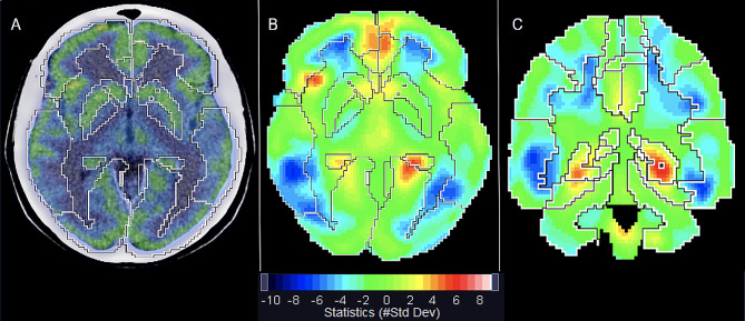
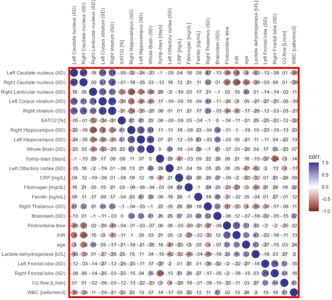
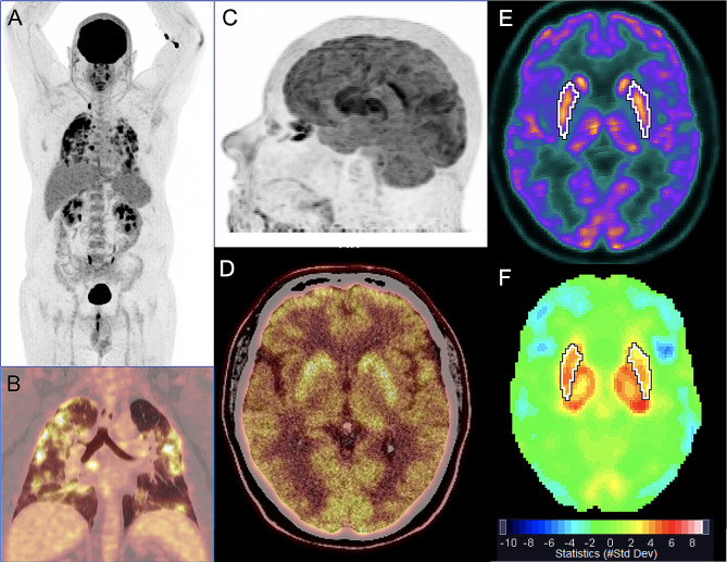
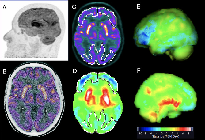
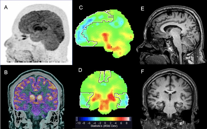
Twenty-three non-intubated patients (13 women; mean age 55.5 ± 12.1 years) hospitalized with positive nasopharyngeal swab test (RT-PCR) for COVID-19, requiring supplemental oxygen and no or mild neurological symptoms were studied. Serum C-reactive protein measured at admission ranged from 6.43 to 189.0 mg/L (mean: 96.9 ± 54.2 mg/L). The mean supplemental oxygen demand was 2.9 ± 1.4 L/min. [F]FDG PET/CT images were acquired with a median of 12 (4-20) days of symptoms. After visual interpretation of the images, semiquantitative analysis of [F]FDG uptake in multiple brain regions was evaluated using dedicated software and the standard deviation (SD) of brain uptake in each region was automatically calculated in comparison with reference values of a normal database. Evolutionarily ancient structures showed positive SD mean values of [F]FDG uptake. Lenticular nuclei were bilaterally hypermetabolic (> 2 SD) in 21/23 (91.3%) patients, and thalamus in 16/23 (69.6%), bilaterally in 11/23 (47.8%). About half of patients showed hypermetabolism in brainstems, 40% in hippocampi, and 30% in cerebellums. In contrast, neocortical regions (frontal, parietal, temporal and occipital lobes) presented negative SD mean values of [F]FDG uptake and hypometabolism (< 2 SD) was observed in up to a third of patients. Associations were found between hypoxia, inflammation, coagulation markers, and [F]FDG uptake in various brain structures.
Brain metabolism is clearly affected during the acute phase of COVID-19 respiratory syndrome in neurologically asymptomatic or oligosymptomatic patients. The most frequent finding is marked hypermetabolism in evolutionary ancient structures such as lenticular nucleus and thalami. Neocortical metabolism was reduced in up to one third of patients, suggesting a redistribution of brain metabolism from the neocortex to evolutionary ancient brain structures in these patients.
新冠病毒病(COVID-19)的神经精神后遗症在疾病慢性或亚急性期出现严重神经症状的患者中已有广泛记录。然而,尚不清楚在疾病急性期早期大脑代谢是否会发生亚临床变化。本研究的目的是识别和量化因COVID-19导致急性呼吸综合征住院且无或仅有轻微神经症状患者的大脑代谢变化。
对23例非插管患者(13例女性;平均年龄55.5±12.1岁)进行了研究,这些患者因COVID-19鼻咽拭子检测(RT-PCR)呈阳性而住院,需要补充氧气且无或仅有轻微神经症状。入院时测得的血清C反应蛋白范围为6.43至189.0mg/L(平均:96.9±54.2mg/L)。平均补充氧气需求量为2.9±1.4L/min。[F]氟代脱氧葡萄糖正电子发射断层扫描/计算机断层扫描([F]FDG PET/CT)图像在症状出现后的中位数为12(4 - 20)天采集。在对图像进行视觉解读后,使用专用软件对多个脑区的[F]FDG摄取进行半定量分析,并与正常数据库的参考值相比自动计算每个区域脑摄取的标准差(SD)。进化上古老的结构显示[F]FDG摄取的SD平均值为正值。21/23(91.3%)的患者双侧豆状核代谢亢进(>2SD),16/23(69.6%)的患者丘脑代谢亢进,其中11/23(47.8%)为双侧。约一半的患者脑干代谢亢进,40%的患者海马体代谢亢进以及30%的患者小脑代谢亢进。相比之下,新皮质区域(额叶、顶叶、颞叶和枕叶)[F]FDG摄取的SD平均值为负值,高达三分之一的患者出现代谢减退(<2SD)。在缺氧、炎症、凝血标志物与不同脑结构的[F]FDG摄取之间发现了相关性。
在神经无症状或症状轻微的患者中,COVID-19呼吸综合征急性期大脑代谢明显受到影响。最常见的发现是豆状核和丘脑等进化上古老的结构出现明显的代谢亢进。高达三分之一的患者新皮质代谢降低,提示这些患者大脑代谢从新皮质重新分布到进化上古老的脑结构。