Xu Danyang, Li Bing, Liu Weixiang, Wei Dan, Long Xiaowu, Huang Tanyu, Lin Hongxin, Cao Kangyang, Zhong Shaonan, Shao Jingjing, Huang Bingsheng, Diao Xian-Fen, Gao Zhenhua
Department of Radiology, The First Affiliated Hospital, Sun Yat-sen University, Guangzhou, China.
Medical AI Lab, School of Biomedical Engineering, Health Science Centre, Shenzhen University, Shenzhen, China.
Quant Imaging Med Surg. 2024 Aug 1;14(8):5420-5433. doi: 10.21037/qims-23-1743. Epub 2024 Jul 12.
Most primary bone tumors are often found in the bone around the knee joint. However, the detection of primary bone tumors on radiographs can be challenging for the inexperienced or junior radiologist. This study aimed to develop a deep learning (DL) model for the detection of primary bone tumors around the knee joint on radiographs.
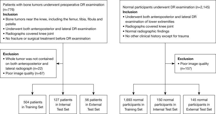
From four tertiary referral centers, we recruited 687 patients diagnosed with bone tumors (including osteosarcoma, chondrosarcoma, giant cell tumor of bone, bone cyst, enchondroma, fibrous dysplasia, etc.; 417 males, 270 females; mean age 22.8±13.2 years) by postoperative pathology or clinical imaging/follow-up, and 1,988 participants with normal bone radiographs (1,152 males, 836 females; mean age 27.9±12.2 years). The dataset was split into a training set for model development, an internal independent and an external test set for model validation. The trained model located bone tumor lesions and then detected tumor patients. Receiver operating characteristic curves and Cohen's kappa coefficient were used for evaluating detection performance. We compared the model's detection performance with that of two junior radiologists in the internal test set using permutation tests.
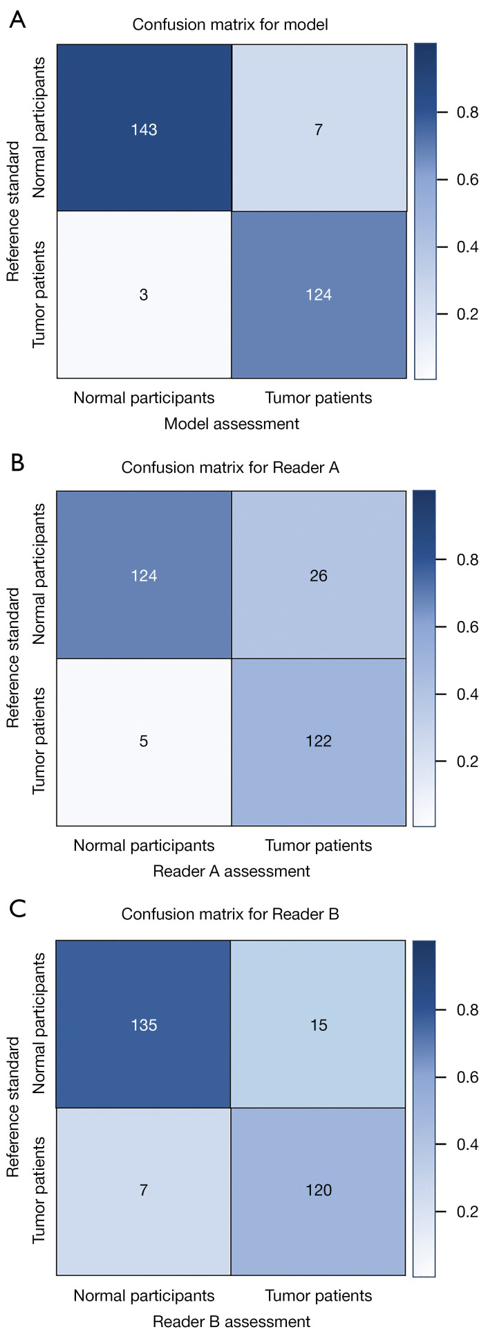
The DL model correctly localized 94.5% and 92.9% bone tumors on radiographs in the internal and external test set, respectively. An accuracy of 0.964/0.920, and an area under the receiver operating characteristic curve (AUC) of 0.981/0.990 in DL detection of bone tumor patients were for the internal and external test set, respectively. Cohen's kappa coefficient of the model in the internal test set was significantly higher than that of the two junior radiologists with 4 and 3 years of experience in musculoskeletal radiology (Model Reader A, 0.927 0.777, P<0.001; Model Reader B, 0.927 0.841, P=0.033).
The DL model achieved good performance in detecting primary bone tumors around the knee joint. This model had better performance than those of junior radiologists, indicating the potential for the detection of bone tumors on radiographs.
大多数原发性骨肿瘤常发生于膝关节周围的骨骼。然而,对于经验不足的初级放射科医生而言,在X线片上检测原发性骨肿瘤具有挑战性。本研究旨在开发一种深度学习(DL)模型,用于在X线片上检测膝关节周围的原发性骨肿瘤。
我们从四个三级转诊中心招募了687例经术后病理或临床影像/随访诊断为骨肿瘤的患者(包括骨肉瘤、软骨肉瘤、骨巨细胞瘤、骨囊肿、内生软骨瘤、骨纤维异常增殖症等;男性417例,女性270例;平均年龄22.8±13.2岁),以及1988例X线片正常的参与者(男性1152例,女性836例;平均年龄27.9±12.2岁)。数据集被分为用于模型开发的训练集、用于模型验证的内部独立测试集和外部测试集。训练后的模型定位骨肿瘤病变,然后检测肿瘤患者。采用受试者操作特征曲线和科恩kappa系数评估检测性能。我们在内部测试集中使用置换检验将模型的检测性能与两名初级放射科医生的检测性能进行比较。
DL模型在内部和外部测试集中分别正确定位了X线片上94.5%和92.9%的骨肿瘤。在DL检测骨肿瘤患者时,内部和外部测试集的准确率分别为0.964/0.920,受试者操作特征曲线下面积(AUC)分别为0.981/0.990。模型在内部测试集中的科恩kappa系数显著高于两名分别具有4年和3年肌肉骨骼放射学经验的初级放射科医生(模型 vs 读者A,0.927 vs 0.777,P<0.001;模型 vs 读者B,0.927 vs 0.841,P=0.033)。
DL模型在检测膝关节周围原发性骨肿瘤方面表现良好。该模型的性能优于初级放射科医生,表明其在X线片上检测骨肿瘤具有潜力。