Pradella Maurice, Baraboo Justin J, Prabhakaran Shyam, Zhao Lihui, Hijaz Tarek, McComb Erin N, Naidich Michelle J, Heckbert Susan R, Nasrallah Ilya M, Bryan R Nick, Passman Rod S, Markl Michael, Greenland Philip
Department of Radiology, Northwestern University Feinberg School of Medicine, Chicago, Illinois, USA.
Department of Radiology, University Hospital Basel, University of Basel, Basel, Switzerland.
J Magn Reson Imaging. 2025 Jan;61(1):276-286. doi: 10.1002/jmri.29349. Epub 2024 Mar 15.
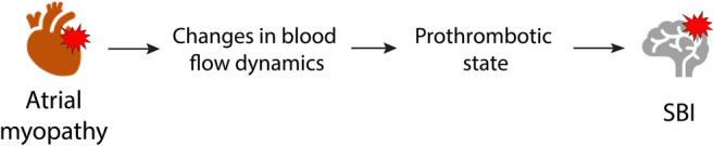
Left atrial (LA) myopathy is thought to be associated with silent brain infarctions (SBI) through changes in blood flow hemodynamics leading to thrombogenesis. 4D-flow MRI enables in-vivo hemodynamic quantification in the left atrium (LA) and LA appendage (LAA).
To determine whether LA and LAA hemodynamic and volumetric parameters are associated with SBI.
Prospective observational study.
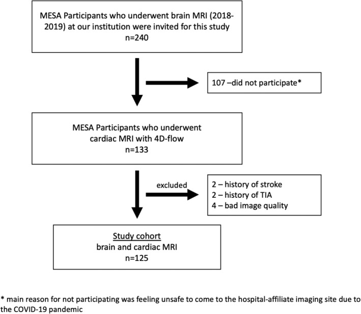
A single-site cohort of 125 Participants of the multiethnic study of atherosclerosis (MESA), mean age: 72.3 ± 7.2 years, 56 men.
FIELD STRENGTH/SEQUENCE: 1.5T. Cardiac MRI: Cine balanced steady state free precession (bSSFP) and 4D-flow sequences. Brain MRI: T1- and T2-weighted SE and FLAIR.
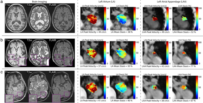
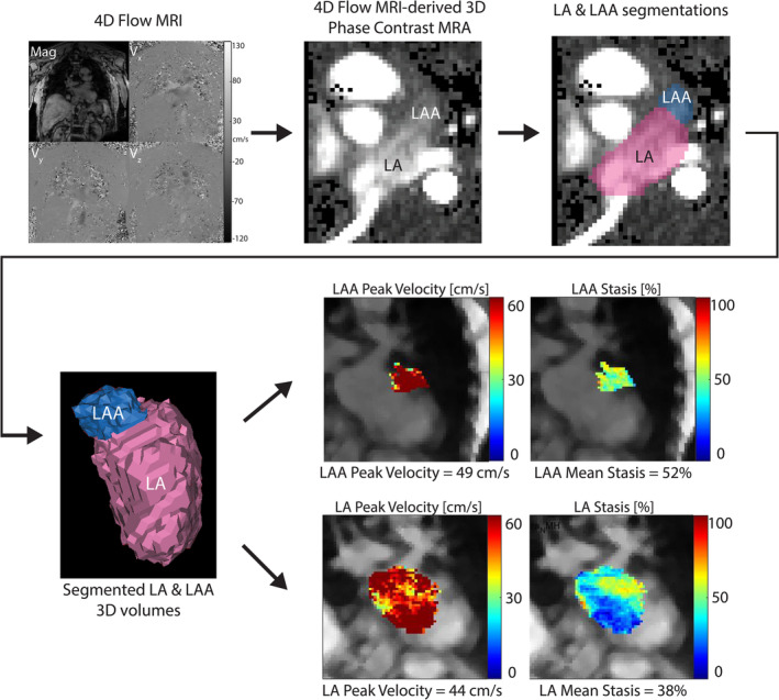
Presence of SBI was determined from brain MRI by neuroradiologists according to routine diagnostic criteria in all participants without a history of stroke based on the MESA database. Minimum and maximum LA volumes and ejection fraction were calculated from bSSFP data. Blood stasis (% of voxels <10 cm/sec) and peak velocity (cm/sec) in the LA and LAA were assessed by a radiologist using an established 4D-flow workflow.
Student's t test, Mann-Whitney U test, one-way ANOVA, chi-square test. Multivariable stepwise logistic regression with automatic forward and backward selection. Significance level P < 0.05.
26 (20.8%) had at least one SBI. After Bonferroni correction, participants with SBI were significantly older and had significantly lower peak velocities in the LAA. In multivariable analyses, age (per 10-years) (odds ratio (OR) = 1.99 (95% confidence interval (CI): 1.30-3.04)) and LAA peak velocity (per cm/sec) (OR = 0.87 (95% CI: 0.81-0.93)) were significantly associated with SBI.
Older age and lower LAA peak velocity were associated with SBI in multivariable analyses whereas volumetric-based measures from cardiac MRI or cardiovascular risk factors were not. Cardiac 4D-flow MRI showed potential to serve as a novel imaging marker for SBI.
3 TECHNICAL EFFICACY: Stage 2.
左心房(LA)肌病被认为通过导致血栓形成的血流动力学变化与无症状性脑梗死(SBI)相关。四维血流磁共振成像(4D-flow MRI)能够对左心房(LA)和左心耳(LAA)进行体内血流动力学定量分析。
确定LA和LAA的血流动力学及容积参数是否与SBI相关。
前瞻性观察性研究。
动脉粥样硬化多族裔研究(MESA)的125名单中心队列参与者,平均年龄:72.3±7.2岁,男性56名。
场强/序列:1.5T。心脏MRI:电影稳态自由进动序列(bSSFP)和4D-flow序列。脑部MRI:T1加权、T2加权自旋回波序列(SE)和液体衰减反转恢复序列(FLAIR)。
由神经放射科医生根据常规诊断标准,通过脑部MRI在所有无卒中病史的参与者中确定SBI的存在情况,数据基于MESA数据库。从bSSFP数据计算LA的最小和最大容积以及射血分数。放射科医生使用既定的4D-flow工作流程评估LA和LAA中的血流淤滞(体素<10 cm/秒的百分比)和峰值速度(cm/秒)。
学生t检验、曼-惠特尼U检验、单因素方差分析、卡方检验。采用自动向前和向后选择的多变量逐步逻辑回归。显著性水平P<0.05。
26名(20.8%)至少有一处SBI。经Bonferroni校正后,患有SBI的参与者年龄显著更大,LAA中的峰值速度显著更低。在多变量分析中,年龄(每10岁)(比值比(OR)=1.99(95%置信区间(CI):1.30 - 3.04))和LAA峰值速度(每厘米/秒)(OR = 0.87(95%CI:0.81 - 0.93))与SBI显著相关。
在多变量分析中,年龄较大和LAA峰值速度较低与SBI相关,而心脏MRI基于容积的测量指标或心血管危险因素则不然。心脏4D-flow MRI显示出作为SBI新型成像标志物的潜力。
3级 技术效能:2级