Davidson Sam J, Saggese Taryn, Krajňáková Jana
Data and Geospatial Intelligence, New Zealand Forest Research Institute (Scion), Christchurch, New Zealand.
Forest Genetics and Biotechnology, New Zealand Forest Research Institute (Scion), Rotorua, New Zealand.
Front Plant Sci. 2024 Mar 1;15:1322920. doi: 10.3389/fpls.2024.1322920. eCollection 2024.
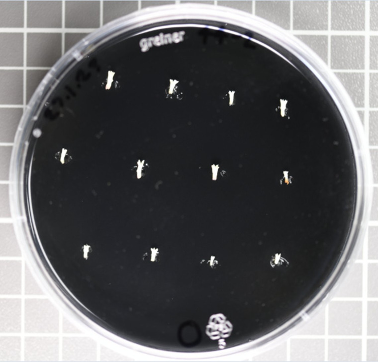
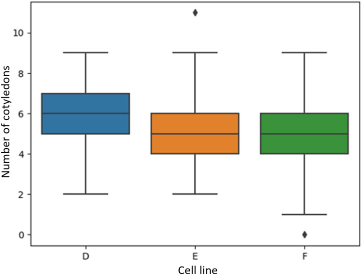
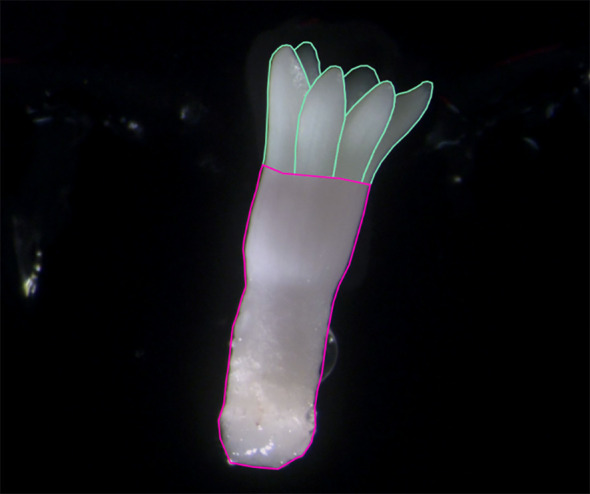
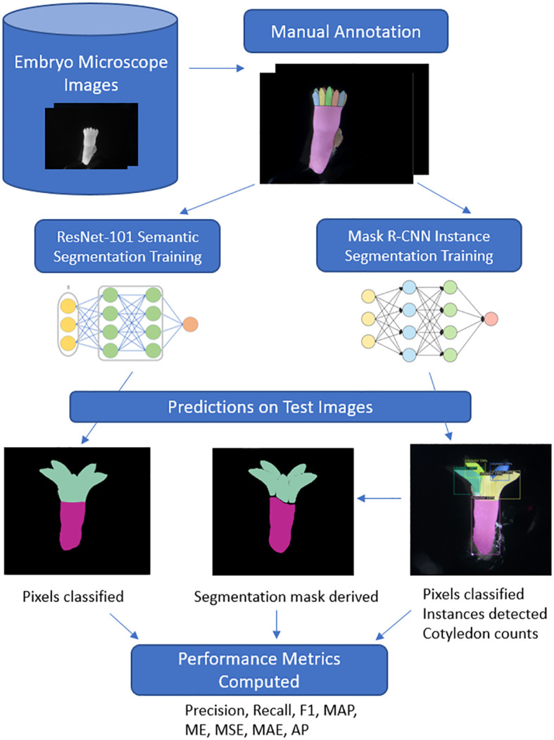
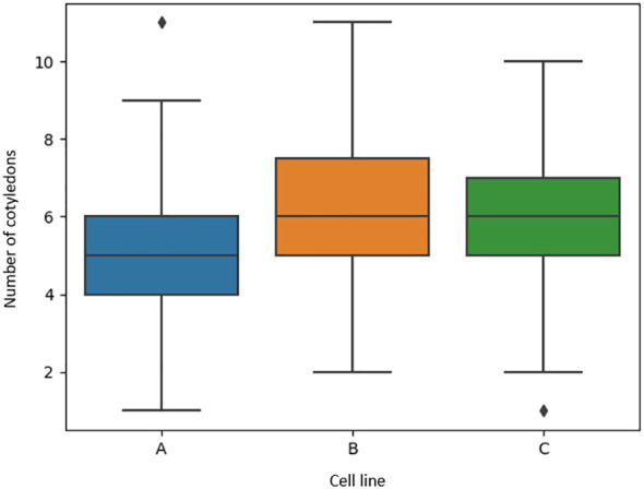
In commercial forestry and large-scale plant propagation, the utilization of artificial intelligence techniques for automated somatic embryo analysis has emerged as a highly valuable tool. Notably, image segmentation plays a key role in the automated assessment of mature somatic embryos. However, to date, the application of Convolutional Neural Networks (CNNs) for segmentation of mature somatic embryos remains unexplored. In this study, we present a novel application of CNNs for delineating mature somatic conifer embryos from background and residual proliferating embryogenic tissue and differentiating various morphological regions within the embryos. A semantic segmentation CNN was trained to assign pixels to cotyledon, hypocotyl, and background regions, while an instance segmentation network was trained to detect individual cotyledons for automated counting. The main dataset comprised 275 high-resolution microscopic images of mature somatic embryos, with 42 images reserved for testing and validation sets. The evaluation of different segmentation methods revealed that semantic segmentation achieved the highest performance averaged across classes, achieving F1 scores of 0.929 and 0.932, with IoU scores of 0.867 and 0.872 for the cotyledon and hypocotyl regions respectively. The instance segmentation approach demonstrated proficiency in accurate detection and counting of the number of cotyledons, as indicated by a mean squared error (MSE) of 0.79 and mean absolute error (MAE) of 0.60. The findings highlight the efficacy of neural network-based methods in accurately segmenting somatic embryos and delineating individual morphological parts, providing additional information compared to previous segmentation techniques. This opens avenues for further analysis, including quantification of morphological characteristics in each region, enabling the identification of features of desirable embryos in large-scale production systems. These advancements contribute to the improvement of automated somatic embryogenesis systems, facilitating efficient and reliable plant propagation for commercial forestry applications.
在商业林业和大规模植物繁殖中,利用人工智能技术进行自动化体细胞胚胎分析已成为一种极具价值的工具。值得注意的是,图像分割在成熟体细胞胚胎的自动化评估中起着关键作用。然而,迄今为止,卷积神经网络(CNN)在成熟体细胞胚胎分割中的应用仍未得到探索。在本研究中,我们展示了CNN的一种新应用,用于从背景和残留的增殖胚性组织中勾勒出成熟的针叶树体细胞胚胎,并区分胚胎内的各种形态区域。训练了一个语义分割CNN,将像素分配到子叶、下胚轴和背景区域,同时训练了一个实例分割网络,以检测单个子叶进行自动计数。主要数据集包括275张成熟体细胞胚胎的高分辨率显微图像,其中42张图像留作测试和验证集。对不同分割方法的评估表明,语义分割在各类别平均性能方面表现最佳,子叶和下胚轴区域的F1分数分别为0.929和0.932,交并比(IoU)分数分别为0.867和0.872。实例分割方法在准确检测和计数子叶数量方面表现出色,平均平方误差(MSE)为0.79,平均绝对误差(MAE)为0.60。研究结果突出了基于神经网络的方法在准确分割体细胞胚胎和描绘单个形态部分方面的有效性,与先前的分割技术相比提供了更多信息。这为进一步分析开辟了途径,包括量化每个区域的形态特征,从而能够在大规模生产系统中识别理想胚胎的特征。这些进展有助于改进自动化体细胞胚胎发生系统,促进商业林业应用中高效可靠的植物繁殖。