• 文献检索
  • 文档翻译
  • 深度研究
  • 学术资讯
  • Suppr Zotero 插件Zotero 插件
  • 邀请有礼
  • 套餐&价格
  • 历史记录
应用&插件
Suppr Zotero 插件Zotero 插件浏览器插件Mac 客户端Windows 客户端微信小程序
定价
高级版会员购买积分包购买API积分包
服务
文献检索文档翻译深度研究API 文档MCP 服务
关于我们
关于 Suppr公司介绍联系我们用户协议隐私条款
关注我们

Suppr 超能文献

核心技术专利:CN118964589B侵权必究
粤ICP备2023148730 号-1Suppr @ 2026

文献检索

告别复杂PubMed语法,用中文像聊天一样搜索,搜遍4000万医学文献。AI智能推荐,让科研检索更轻松。

立即免费搜索

文件翻译

保留排版,准确专业,支持PDF/Word/PPT等文件格式,支持 12+语言互译。

免费翻译文档

深度研究

AI帮你快速写综述,25分钟生成高质量综述,智能提取关键信息,辅助科研写作。

立即免费体验

利用深度学习进行图像生成插值和恢复以改进3D组织映射

Generative interpolation and restoration of images using deep learning for improved 3D tissue mapping.

作者信息

Joshi Saurabh, Forjaz André, Han Kyu Sang, Shen Yu, Queiroga Vasco, Xenes Daniel, Matelsk Jordan, Wester Brock, Barrutia Arrate Munoz, Kiemen Ashley L, Wu Pei-Hsun, Wirtz Denis

机构信息

Department of Chemical & Biomolecular Engineering, Johns Hopkins University, Baltimore MD.

The Johns Hopkins Institute for NanoBioTechnology, Johns Hopkins University, Baltimore, MD.

出版信息

bioRxiv. 2024 Mar 28:2024.03.07.583909. doi: 10.1101/2024.03.07.583909.

DOI:10.1101/2024.03.07.583909
PMID:38496512
原文链接:https://pmc.ncbi.nlm.nih.gov/articles/PMC10942457/
Abstract

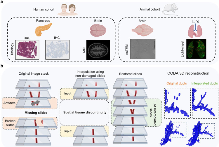
The development of novel imaging platforms has improved our ability to collect and analyze large three-dimensional (3D) biological imaging datasets. Advances in computing have led to an ability to extract complex spatial information from these data, such as the composition, morphology, and interactions of multi-cellular structures, rare events, and integration of multi-modal features combining anatomical, molecular, and transcriptomic (among other) information. Yet, the accuracy of these quantitative results is intrinsically limited by the quality of the input images, which can contain missing or damaged regions, or can be of poor resolution due to mechanical, temporal, or financial constraints. In applications ranging from intact imaging (e.g. light-sheet microscopy and magnetic resonance imaging) to sectioning based platforms (e.g. serial histology and serial section transmission electron microscopy), the quality and resolution of imaging data has become paramount. Here, we address these challenges by leveraging frame interpolation for large image motion (FILM), a generative AI model originally developed for temporal interpolation, for spatial interpolation of a range of 3D image types. Comparative analysis demonstrates the superiority of FILM over traditional linear interpolation to produce functional synthetic images, due to its ability to better preserve biological information including microanatomical features and cell counts, as well as image quality, such as contrast, variance, and luminance. FILM repairs tissue damages in images and reduces stitching artifacts. We show that FILM can decrease imaging time by synthesizing skipped images. We demonstrate the versatility of our method with a wide range of imaging modalities (histology, tissue-clearing/light-sheet microscopy, magnetic resonance imaging, serial section transmission electron microscopy), species (human, mouse), healthy and diseased tissues (pancreas, lung, brain), staining techniques (IHC, H&E), and pixel resolutions (8 nm, 2 μm, 1mm). Overall, we demonstrate the potential of generative AI in improving the resolution, throughput, and quality of biological image datasets, enabling improved 3D imaging.

摘要

新型成像平台的发展提高了我们收集和分析大型三维(3D)生物成像数据集的能力。计算技术的进步使我们有能力从这些数据中提取复杂的空间信息,例如多细胞结构的组成、形态和相互作用、罕见事件,以及结合解剖学、分子和转录组学(以及其他)信息的多模态特征的整合。然而,这些定量结果的准确性本质上受到输入图像质量的限制,输入图像可能包含缺失或受损区域,或者由于机械、时间或经济限制而分辨率较低。在从完整成像(如光片显微镜和磁共振成像)到基于切片的平台(如连续组织学和连续切片透射电子显微镜)的各种应用中,成像数据的质量和分辨率已变得至关重要。在这里,我们通过利用用于大图像运动的帧插值(FILM)来应对这些挑战,FILM是一种最初为时间插值而开发的生成式人工智能模型,用于一系列3D图像类型的空间插值。对比分析表明,FILM在生成功能合成图像方面优于传统线性插值,因为它能够更好地保留包括微观解剖特征和细胞计数在内的生物信息,以及图像质量,如对比度、方差和亮度。FILM修复图像中的组织损伤并减少拼接伪影。我们表明,FILM可以通过合成跳过的图像来减少成像时间。我们用广泛的成像模态(组织学、组织清除/光片显微镜、磁共振成像、连续切片透射电子显微镜)、物种(人类、小鼠)、健康和患病组织(胰腺、肺、脑)、染色技术(免疫组化、苏木精和伊红染色)和像素分辨率(8纳米、2微米、1毫米)证明了我们方法的通用性。总体而言,我们展示了生成式人工智能在提高生物图像数据集的分辨率、通量和质量方面的潜力,从而实现改进的3D成像。

https://cdn.ncbi.nlm.nih.gov/pmc/blobs/621c/11005578/06f06bc15819/nihpp-2024.03.07.583909v3-f0005.jpg
https://cdn.ncbi.nlm.nih.gov/pmc/blobs/621c/11005578/07c3e345ec9f/nihpp-2024.03.07.583909v3-f0006.jpg
https://cdn.ncbi.nlm.nih.gov/pmc/blobs/621c/11005578/fa874a856c00/nihpp-2024.03.07.583909v3-f0001.jpg
https://cdn.ncbi.nlm.nih.gov/pmc/blobs/621c/11005578/1b670d1ad460/nihpp-2024.03.07.583909v3-f0002.jpg
https://cdn.ncbi.nlm.nih.gov/pmc/blobs/621c/11005578/d61bc1d2f1e3/nihpp-2024.03.07.583909v3-f0003.jpg
https://cdn.ncbi.nlm.nih.gov/pmc/blobs/621c/11005578/c5974848d682/nihpp-2024.03.07.583909v3-f0004.jpg
https://cdn.ncbi.nlm.nih.gov/pmc/blobs/621c/11005578/06f06bc15819/nihpp-2024.03.07.583909v3-f0005.jpg

相似文献

1
Generative interpolation and restoration of images using deep learning for improved 3D tissue mapping.利用深度学习进行图像生成插值和恢复以改进3D组织映射
bioRxiv. 2024 Mar 28:2024.03.07.583909. doi: 10.1101/2024.03.07.583909.
2
InterpolAI: deep learning-based optical flow interpolation and restoration of biomedical images for improved 3D tissue mapping.InterpolAI:基于深度学习的光流插值与生物医学图像恢复,用于改进三维组织映射
Nat Methods. 2025 May 28. doi: 10.1038/s41592-025-02712-4.
3
Leveraging a foundation model zoo for cell similarity search in oncological microscopy across devices.利用基础模型库进行跨设备肿瘤显微镜检查中的细胞相似性搜索。
Front Oncol. 2025 Jun 18;15:1480384. doi: 10.3389/fonc.2025.1480384. eCollection 2025.
4
Linking transcriptome and morphology in bone cells at cellular resolution with generative AI.利用生成式人工智能在细胞分辨率下将骨细胞中的转录组与形态学联系起来。
J Bone Miner Res. 2024 Dec 31;40(1):20-26. doi: 10.1093/jbmr/zjae151.
5
Artificial intelligence for diagnosing exudative age-related macular degeneration.人工智能在渗出性年龄相关性黄斑变性诊断中的应用。
Cochrane Database Syst Rev. 2024 Oct 17;10(10):CD015522. doi: 10.1002/14651858.CD015522.pub2.
6
Management of urinary stones by experts in stone disease (ESD 2025).结石病专家对尿路结石的管理(2025年结石病专家共识)
Arch Ital Urol Androl. 2025 Jun 30;97(2):14085. doi: 10.4081/aiua.2025.14085.
7
Novel application of metabolic imaging of early embryos using a light-sheet on-a-chip device: a proof-of-concept study.使用片上光片装置对早期胚胎进行代谢成像的新应用:一项概念验证研究。
Hum Reprod. 2025 Jan 1;40(1):41-55. doi: 10.1093/humrep/deae249.
8
Influence of early through late fusion on pancreas segmentation from imperfectly registered multimodal magnetic resonance imaging.早期至晚期融合对来自配准不完善的多模态磁共振成像的胰腺分割的影响。
J Med Imaging (Bellingham). 2025 Mar;12(2):024008. doi: 10.1117/1.JMI.12.2.024008. Epub 2025 Apr 26.
9
Magnetic resonance perfusion for differentiating low-grade from high-grade gliomas at first presentation.首次就诊时磁共振灌注成像用于鉴别低级别与高级别胶质瘤
Cochrane Database Syst Rev. 2018 Jan 22;1(1):CD011551. doi: 10.1002/14651858.CD011551.pub2.
10
Interventions for central serous chorioretinopathy: a network meta-analysis.中心性浆液性脉络膜视网膜病变的干预措施:一项网状Meta分析
Cochrane Database Syst Rev. 2025 Jun 16;6(6):CD011841. doi: 10.1002/14651858.CD011841.pub3.

引用本文的文献

1
COEXIST: Coordinated single-cell integration of serial multiplexed tissue images.COEXIST:连续多重组织图像的协调单细胞整合
PLoS Comput Biol. 2025 Aug 5;21(8):e1013325. doi: 10.1371/journal.pcbi.1013325. eCollection 2025 Aug.

本文引用的文献

1
PanIN and CAF transitions in pancreatic carcinogenesis revealed with spatial data integration.通过空间数据整合揭示胰腺癌发生过程中的胰腺上皮内瘤变(PanIN)和癌症相关成纤维细胞(CAF)转变
Cell Syst. 2024 Aug 21;15(8):753-769.e5. doi: 10.1016/j.cels.2024.07.001. Epub 2024 Aug 7.
2
SynCLay: Interactive synthesis of histology images from bespoke cellular layouts.SynCLay:根据定制的细胞排布交互式合成组织学图像。
Med Image Anal. 2024 Jan;91:102995. doi: 10.1016/j.media.2023.102995. Epub 2023 Oct 11.
3
Zero-Shot Medical Image Translation via Frequency-Guided Diffusion Models.
基于频域引导扩散模型的零样本医学图像翻译。
IEEE Trans Med Imaging. 2024 Mar;43(3):980-993. doi: 10.1109/TMI.2023.3325703. Epub 2024 Mar 5.
4
Diffusion models in medical imaging: A comprehensive survey.扩散模型在医学成像中的应用:全面综述。
Med Image Anal. 2023 Aug;88:102846. doi: 10.1016/j.media.2023.102846. Epub 2023 May 23.
5
Spatial Transcriptomics of Intraductal Papillary Mucinous Neoplasms of the Pancreas Identifies NKX6-2 as a Driver of Gastric Differentiation and Indolent Biological Potential.胰腺导管内乳头状黏液性肿瘤的空间转录组学鉴定 NKX6-2 为胃分化和惰性生物学潜能的驱动因素。
Cancer Discov. 2023 Aug 4;13(8):1844-1861. doi: 10.1158/2159-8290.CD-22-1200.
6
Light-sheets and smart microscopy, an exciting future is dawning.光片和智能显微镜,一个激动人心的未来正在到来。
Commun Biol. 2023 May 9;6(1):502. doi: 10.1038/s42003-023-04857-4.
7
Denoising diffusion probabilistic models for 3D medical image generation.基于去噪扩散概率模型的三维医学图像生成。
Sci Rep. 2023 May 5;13(1):7303. doi: 10.1038/s41598-023-34341-2.
8
Deep learning-enabled virtual histological staining of biological samples.基于深度学习的生物样本虚拟组织学染色
Light Sci Appl. 2023 Mar 3;12(1):57. doi: 10.1038/s41377-023-01104-7.
9
Unstained Tissue Imaging and Virtual Hematoxylin and Eosin Staining of Histologic Whole Slide Images.组织切片全视野图像的未染色组织成像和虚拟苏木精-伊红染色。
Lab Invest. 2023 May;103(5):100070. doi: 10.1016/j.labinv.2023.100070. Epub 2023 Jan 25.
10
Multiplexed 3D atlas of state transitions and immune interaction in colorectal cancer.多指标 3D 图谱描绘结直肠癌中状态转变和免疫相互作用。
Cell. 2023 Jan 19;186(2):363-381.e19. doi: 10.1016/j.cell.2022.12.028.