• 文献检索
  • 文档翻译
  • 深度研究
  • 学术资讯
  • Suppr Zotero 插件Zotero 插件
  • 邀请有礼
  • 套餐&价格
  • 历史记录
应用&插件
Suppr Zotero 插件Zotero 插件浏览器插件Mac 客户端Windows 客户端微信小程序
定价
高级版会员购买积分包购买API积分包
服务
文献检索文档翻译深度研究API 文档MCP 服务
关于我们
关于 Suppr公司介绍联系我们用户协议隐私条款
关注我们

Suppr 超能文献

核心技术专利:CN118964589B侵权必究
粤ICP备2023148730 号-1Suppr @ 2026

文献检索

告别复杂PubMed语法,用中文像聊天一样搜索,搜遍4000万医学文献。AI智能推荐,让科研检索更轻松。

立即免费搜索

文件翻译

保留排版,准确专业,支持PDF/Word/PPT等文件格式,支持 12+语言互译。

免费翻译文档

深度研究

AI帮你快速写综述,25分钟生成高质量综述,智能提取关键信息,辅助科研写作。

立即免费体验

基于环境感应染料荧光寿命的活细胞生物传感器。

Live-cell biosensors based on the fluorescence lifetime of environment-sensing dyes.

机构信息

Department of Pharmacology, University of North Carolina at Chapel Hill, Chapel Hill, NC 27599, USA.

Laboratory for Fluorescence Dynamics, Department of Biomedical Engineering, University of California at Irvine, Irvine, CA 92617, USA.

出版信息

Cell Rep Methods. 2024 Mar 25;4(3):100734. doi: 10.1016/j.crmeth.2024.100734. Epub 2024 Mar 18.

DOI:10.1016/j.crmeth.2024.100734
PMID:38503289
原文链接:https://pmc.ncbi.nlm.nih.gov/articles/PMC10985238/
Abstract

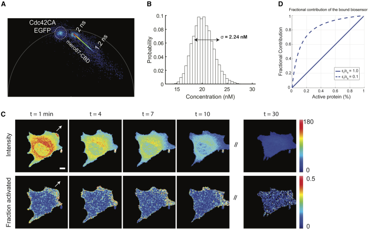
In this work, we examine the use of environment-sensitive fluorescent dyes in fluorescence lifetime imaging microscopy (FLIM) biosensors. We screened merocyanine dyes to find an optimal combination of environment-induced lifetime changes, photostability, and brightness at wavelengths suitable for live-cell imaging. FLIM was used to monitor a biosensor reporting conformational changes of endogenous Cdc42 in living cells. The ability to quantify activity using phasor analysis of a single fluorophore (e.g., rather than ratio imaging) eliminated potential artifacts. We leveraged these properties to determine specific concentrations of activated Cdc42 across the cell.

摘要

在这项工作中,我们研究了环境敏感荧光染料在荧光寿命成像显微镜(FLIM)生物传感器中的应用。我们筛选了甲川花菁染料,以找到在适合活细胞成像的波长下,环境诱导寿命变化、光稳定性和亮度的最佳组合。FLIM 用于监测生物传感器报告活细胞内内源性 Cdc42 构象变化。使用单个荧光团的相分析(例如,而不是比率成像)来定量活性的能力消除了潜在的伪影。我们利用这些特性来确定细胞内特定浓度的激活 Cdc42。

https://cdn.ncbi.nlm.nih.gov/pmc/blobs/d82c/10985238/e9ca81296335/fx12.jpg
https://cdn.ncbi.nlm.nih.gov/pmc/blobs/d82c/10985238/dfdc82c5ec20/fx1.jpg
https://cdn.ncbi.nlm.nih.gov/pmc/blobs/d82c/10985238/e74e9ed15918/gr1.jpg
https://cdn.ncbi.nlm.nih.gov/pmc/blobs/d82c/10985238/a3be25861050/gr2.jpg
https://cdn.ncbi.nlm.nih.gov/pmc/blobs/d82c/10985238/0d878aad7437/gr3.jpg
https://cdn.ncbi.nlm.nih.gov/pmc/blobs/d82c/10985238/0ef6901e4e22/gr4.jpg
https://cdn.ncbi.nlm.nih.gov/pmc/blobs/d82c/10985238/f37fb5847324/fx2.jpg
https://cdn.ncbi.nlm.nih.gov/pmc/blobs/d82c/10985238/eded6c6e2df2/fx3.jpg
https://cdn.ncbi.nlm.nih.gov/pmc/blobs/d82c/10985238/7fb3650204dc/fx4.jpg
https://cdn.ncbi.nlm.nih.gov/pmc/blobs/d82c/10985238/9385735116cf/fx5.jpg
https://cdn.ncbi.nlm.nih.gov/pmc/blobs/d82c/10985238/b6f95c67731a/fx6.jpg
https://cdn.ncbi.nlm.nih.gov/pmc/blobs/d82c/10985238/1498991ddabb/fx7.jpg
https://cdn.ncbi.nlm.nih.gov/pmc/blobs/d82c/10985238/293a05cd522c/fx8.jpg
https://cdn.ncbi.nlm.nih.gov/pmc/blobs/d82c/10985238/37a6b0742a0f/fx9.jpg
https://cdn.ncbi.nlm.nih.gov/pmc/blobs/d82c/10985238/da2db3a56d03/fx10.jpg
https://cdn.ncbi.nlm.nih.gov/pmc/blobs/d82c/10985238/fa292b578a4b/fx11.jpg
https://cdn.ncbi.nlm.nih.gov/pmc/blobs/d82c/10985238/e9ca81296335/fx12.jpg

相似文献

1
Live-cell biosensors based on the fluorescence lifetime of environment-sensing dyes.基于环境感应染料荧光寿命的活细胞生物传感器。
Cell Rep Methods. 2024 Mar 25;4(3):100734. doi: 10.1016/j.crmeth.2024.100734. Epub 2024 Mar 18.
2
Environment-sensing merocyanine dyes for live cell imaging applications.用于活细胞成像应用的环境感应型次甲基菁染料。
Bioconjug Chem. 2013 Feb 20;24(2):215-23. doi: 10.1021/bc3005073. Epub 2013 Jan 16.
3
Solvent-sensitive dyes to report protein conformational changes in living cells.用于报告活细胞中蛋白质构象变化的溶剂敏感染料。
J Am Chem Soc. 2003 Apr 9;125(14):4132-45. doi: 10.1021/ja0290882.
4
Biosensor Förster resonance energy transfer detection by the phasor approach to fluorescence lifetime imaging microscopy.荧光寿命成像显微镜中通过相位荧光寿命检测生物传感器的Förster 共振能量转移。
Microsc Res Tech. 2012 Mar;75(3):271-81. doi: 10.1002/jemt.21054. Epub 2011 Aug 19.
5
PIE-FLIM Measurements of Two Different FRET-Based Biosensor Activities in the Same Living Cells.在同一活细胞中对两种不同基于荧光共振能量转移的生物传感器活性进行的受激拉曼散射显微镜测量。
Biophys J. 2020 Apr 21;118(8):1820-1829. doi: 10.1016/j.bpj.2020.03.003. Epub 2020 Mar 10.
6
Phasor Analysis of Fluorescence Lifetime Enables Quantitative Multiplexed Molecular Imaging of Three Probes.荧光寿命相分析实现三种探针的定量多重分子成像
Anal Chem. 2022 Oct 18;94(41):14185-14194. doi: 10.1021/acs.analchem.2c02149. Epub 2022 Oct 3.
7
Phasor S-FLIM: a new paradigm for fast and robust spectral fluorescence lifetime imaging.相衬 S-FLIM:一种快速稳健的光谱荧光寿命成像新范例。
Nat Methods. 2021 May;18(5):542-550. doi: 10.1038/s41592-021-01108-4. Epub 2021 Apr 15.
8
Optimization of Advanced Live-Cell Imaging through Red/Near-Infrared Dye Labeling and Fluorescence Lifetime-Based Strategies.通过红/近红外染料标记和荧光寿命的策略优化高级活细胞成像。
Int J Mol Sci. 2021 Oct 14;22(20):11092. doi: 10.3390/ijms222011092.
9
Solvatochromic and Fluorogenic Dyes as Environment-Sensitive Probes: Design and Biological Applications.溶剂化变色和荧光染料作为环境敏感探针:设计与生物学应用。
Acc Chem Res. 2017 Feb 21;50(2):366-375. doi: 10.1021/acs.accounts.6b00517. Epub 2017 Jan 9.
10
Quantitative real-time imaging of intracellular FRET biosensor dynamics using rapid multi-beam confocal FLIM.利用快速多光束共焦 FLIM 对细胞内 FRET 生物传感器动态进行定量实时成像。
Sci Rep. 2020 Mar 20;10(1):5146. doi: 10.1038/s41598-020-61478-1.

本文引用的文献

1
The Phasor Plot: A Universal Circle to Advance Fluorescence Lifetime Analysis and Interpretation.相量图:推进荧光寿命分析和解释的通用圆。
Annu Rev Biophys. 2021 May 6;50:575-593. doi: 10.1146/annurev-biophys-062920-063631.
2
Multiplexed GTPase and GEF biosensor imaging enables network connectivity analysis.多重 GTPase 和 GEF 生物传感器成像可实现网络连通性分析。
Nat Chem Biol. 2020 Aug;16(8):826-833. doi: 10.1038/s41589-020-0542-9. Epub 2020 May 18.
3
Fluorescence lifetime imaging microscopy: fundamentals and advances in instrumentation, analysis, and applications.
荧光寿命成像显微镜:仪器、分析和应用的基本原理及进展。
J Biomed Opt. 2020 May;25(7):1-43. doi: 10.1117/1.JBO.25.7.071203.
4
Membrane-Permeant, Environment-Sensitive Dyes Generate Biosensors within Living Cells.膜透性、环境敏感染料在活细胞内生成生物传感器。
J Am Chem Soc. 2019 May 8;141(18):7275-7282. doi: 10.1021/jacs.8b09841. Epub 2019 Apr 23.
5
Genetically Encoded Fluorescent Biosensors Illuminate the Spatiotemporal Regulation of Signaling Networks.基因编码荧光生物传感器照亮信号网络的时空调控。
Chem Rev. 2018 Dec 26;118(24):11707-11794. doi: 10.1021/acs.chemrev.8b00333. Epub 2018 Dec 14.
6
Monitoring intracellular nanomolar calcium using fluorescence lifetime imaging.利用荧光寿命成像监测细胞内纳摩尔级钙离子。
Nat Protoc. 2018 Mar;13(3):581-597. doi: 10.1038/nprot.2017.154. Epub 2018 Feb 22.
7
Solvatochromic and Fluorogenic Dyes as Environment-Sensitive Probes: Design and Biological Applications.溶剂化变色和荧光染料作为环境敏感探针:设计与生物学应用。
Acc Chem Res. 2017 Feb 21;50(2):366-375. doi: 10.1021/acs.accounts.6b00517. Epub 2017 Jan 9.
8
Ratiometric Imaging Using a Single Dye Enables Simultaneous Visualization of Rac1 and Cdc42 Activation.使用单一染料的比率成像可实现Rac1和Cdc42激活的同时可视化。
J Am Chem Soc. 2016 Mar 2;138(8):2571-5. doi: 10.1021/jacs.5b09764. Epub 2016 Feb 19.
9
Separating NADH and NADPH fluorescence in live cells and tissues using FLIM.利用荧光寿命成像技术(FLIM)区分活细胞和组织中的NADH和NADPH荧光。
Nat Commun. 2014 May 29;5:3936. doi: 10.1038/ncomms4936.
10
Cellular incorporation of unnatural amino acids and bioorthogonal labeling of proteins.非天然氨基酸的细胞掺入与蛋白质的生物正交标记
Chem Rev. 2014 May 14;114(9):4764-806. doi: 10.1021/cr400355w. Epub 2014 Mar 21.